Spinal Decompression Research
NYC Spinal Decompression Specialist DRX 9000 (212) 645-8151

DISTRACTIVE VERTEBRAL AXIAL DECOMPRESSION: PATIENT OUTCOME STUDY
by Vicki Ludwin-Brown, CPHQ, RCP, RRT
8/15/2004
Summary: The outcomes of distractive vertebral axial decompression therapy for patients with low back pain from various causes was reported by Gose, Naguszewski and Naguszewski in "Vertebral axial decompression therapy for pain associated with herniated or degenerated disc or facet syndrome: An Outcome Study" - Neurological Research, Vol. 20, No. 3, April 1998.
"Data was collected from twenty-two medical centers for patients who received VAX-D therapy for low pain, which was sometimes accompanied by referred leg pain. Only patients who received at least ten sessions and had a diagnosis of herniated disc, degenerative disc, or facet syndrome, which were confirmed by diagnostic imaging, were included in this study; a total of 778 cases. The data contained the patients' quantitative assessments of their own pain, mobility and ability to carry out the usual activities of daily living. The treatment was successful in 72% of the 778 cases, when success was defined as a reduction in pain to 0 or 1, on a 0-5 scale. Improvements in mobility and activities of daily living correlated strongly with pain reduction."
In an effort to determine the outcomes of our own patients in this community, we studied our first 51 back patients at ReNew Rehab and Back 'N Balance. Our outcomes were similar to those reported in the aforementioned article. Additionally, our outcomes were similar with the first 33 patients as compared to the larger sample size of 49 patients. As part of the program evaluation process, The UR committee discussed the program strengths and weaknesses (lack of community and physician knowledge of our application).
| # | % | ||
| Great improvement | 25 | ||
| Good improvement |
|
||
| Some improvement |
|
||
| No improvement |
|
||
| Total |
|
| Satisfaction |
|
|||
| Very Satisfied |
|
|||
| Satisfied | 14 | |||
| Not satisfied |
|
|||
| Total | 49 |
The Utilization Review Committee reviewed all chronic low back pain cases for appropriateness and efficacy of services related to the low back pain program, Distractive Vertebral Axial Decompression.
The quality improvement program related to Distractive Vertebral Axial Decompression Therapy that has been implemented at Back `N Balance and formerly at ReNew Rehab includes the following components:
* Equipment Quality Control (VAX-D was used for all patients)
* Quarterly Equipment Calibration per manufacturer's guidelines
* Treatment waveform documentation per manufacturer's guidelines
* Operator Quality Control * Certification of operator * Training Program with Preceptor (20 sessions)
* Clinical Assessment * Physician equipment certification * Physician patient assessment per Manufacturer's Protocol (VAX-D) * Therapy sessions per Protocol * Physician re-assessment per Protocol * Physician assessment mid-term if lack of progress * Case review by Medical Director of all individual cases * Medical Director review of aggregated results
Period Reviewed: December 2002-August 2004
Utilization Review (UR) Committee members: Vicki Brown, C.P.H.Q., Program Coordinator, Certified VAX-D technician Patti Boos, Records Keeper and Office Manager Glenn A. Thomas, M.D. Denise Endly, R.N.
Process: All patient charts (49) were reviewed for the following criteria:
(1) Adherence to Protocol (24 treatments - 5X week X 4 weeks then 1X/week X 4 weeks) (2) Efficacy based on pain scale and/or Oswestry Disability index start and end of therapy (3) Evidence of physician review (4) Patient satisfaction
Data Process:
-
- Data collection * Pain scale, Oswestry, H&P at initial visit (Back `N Balance, formerly ReNew Rehab) * SF-12 at beginning and >1 month post care (vendor/QHR) and follow up calls before
- each utilization review committee meeting * Satisfaction Survey at end of therapy (Back `N Balance/formerly ReNew Rehab) * Daily pain and patient comments documented in technician progress reports * Physician evaluations of patient (2-3: @ start, mid-term, completion of therapy)
- Data Entry * Access database for individual entries - by Program Coordinator or Office Manager
- Case review * Physician and Program Coordinator with physician reassessments
- Data aggregation in Excel database - facilitated by Program Coordinator and assistants * Reviewed by UR Committee
Quality monitoring is done concurrently (daily progress notes) and retrospectively in aggregate (by UR committee). Quality control is completed quarterly.
Results:
Of the 51 total patients, 49 had at least 10 sessions. The reasons for (7) patients not completing the usual treatment protocol of 24 treatments included:
- Patient improvement or plateau before 24 treatments (2 patients)
- Patient lack of compliance with protocol (1)
- Patient inability to pay for further treatments (2 patients)
- Patient discomfort with treatment - Inability to lay prone for 30 minutes or pain getting off the table (2 patients)
49 charts of all patients receiving 10 or more treatments were reviewed for the following:
(1) Appropriateness of care:
100% of the charts demonstrated that the protocol was followed: * Patient selection was appropriate per protocol * Therapy proceeded per VAX-D Protocol * Each case was reviewed by the physician
(2) Efficacy of care:
Of the 49 patients treated with > 10 treatments of vertebral axial decompression therapy, 39/49 or 80% of the charts reviewed demonstrated that the patient improved to minimal or no pain post therapy (0-1 on a pain scale of 1-5*).
* No improvement (4/49) =8% (no change in pain scale rating)* Some improvement (10/49) = 20.5% (change of 1 increment in pain scale)* Good improvement (10/49) = 20.5% (change of 2 increments in pain scale) * Great improvement (25/49) = 51% (change of 3 increments in pain scale)
*Pain Rating scale (Oswestry): (0=no pain, 1=mild pain, 2=moderate pain, 3=fairly severe pain, 4=very severe pain, 5=worst imaginable)
(3) Patient Satisfaction:
43/49 or 88% patients were satisfied or very satisfied with therapy. * 6/49 or 12% of patients were not satisfied * 14/49 or 29% of patients were satisfied * 29/49 or 59% of patients were very satisfied
(4) Additional findings:
a. Patient history of previous therapy/back surgery prior to vertebral axial decompression therapy * 28/49 patients (57%) had prior Physical Therapy * 30/49 patients (61%) had prior chiropractic care * 28/49 patients (57%) had prior injections for pain * 13/49 patients (27%) had prior back surgery b. Average # of treatments per patient = 25 c. Patients ranged in ages from 18 to 81 - Average age was 51 years d. Average # of years history of back pain- 8 years - Range from .3 to 30 year history e. Diagnoses were as follows:* 48/49 patients (98%) had lumbar bulging, herniated, protruding, or extruded discs
varying in size from 2mm to 15mm (per MRI report) * 36/49 patients (73%) had concomitant degenerative disc disease * 36/49 patients (73%) had multilevel issues or multiple diagnoses * 35/49 patients (71%) complained of sciatica
f. Surgery after course of therapy = 1/49 or 2%
© Vicki Ludwin-Brown, all rights reserved. Back 'n Balance, 5876 Owens Avenue, Suite 100, Carlsbad, CA. 92008 (760) 602-3134 BackSanDiego.com [email protected]
VAX-D® PROTOCOL and Guidelines
Therapeutic Indications for VAX-D
- Low back pain, unilateral or bilateral, with or without leg pain associated with discogenic lesions.
- Patients with neurologic deficits
- Post surgical patients with failed back syndrome without retained hardware
Contraindications and Potential risks for VAX-D Patients
- Primary or metastatic neoplasm in the spine
- Vertebral fracture (recent)
- Cauda equine syndrome
- Unstable spondylolisthesis (eg Pars Defects)
- Severe osteoporosis - plain films show greater than 45% bone loss or less than 2.5 standard deviations below the mean on a DEXA test
- Severe or unstable medical disorder
- Significant shoulder injury - rotator cuff tear
- Pregnancy
- Ankylosing spondylitis
- Arthodesis with retained hardware
- Abdominal aortic aneurysm
- Spinal infections including osteomyelitis and septic discitis
- Severe osseous stenosis
- Hemangioma that invades the endplate, greater than 1cm in size that may compromise the vertebral integrity or that may represent a risk of bleeding
- Pelvic or abdominal cancer
Vertebral Axial Decompression Therapy for Pain Associated With Herniated or Degenerated Discs or Facet Syndrome: An Outcome Study
Earl E. Gose, William K. Naguszewski* and Robert K. Naguszewski*
Department of Bioengineering, University of Iliinois at Chicago, Chicago, IL. USA *Coosa Medical Group, Rome, Georgia, USA
The outcomes of vertebral axial decompression (VAX-D) therapy for patients with low back pain from various causes are reported. Data was collected from twenty-two medical centers for patients who received VAX-D therapy for low back pain, which was sometimes accompanied by referred leg pain. Only patients who received at least ten sessions and had a diagnosis of herniated disc, degenerative disc, or facet syndrome, which were confirmed by diagnostic imaging, were included in this study; a total of 778 cases. The average time between the initial onset of symptoms and the beginning of this therapy was 40 months, and it was four months or more in 83% of the cases. The data contained the patients' quantitative assessments of their own pain, mobility, and ability to carry out the usual 'activities of daily living'. The treatment was successful in 71% of the 778 cases, when success was defined as a reduction in pain to 0 or 1, on a 0 to 5 scale. Improvements in mobility and activities of daily living correlated strongly with pain reduction. The causes of back pain and their relationship to this therapy are also discussed. [Neurol Res 1998; 20: 186-190].
Keywords: Low back pain; herniated disc
INTRODUCTION
For most patients, the cause or causes of persistent low back pain remains poorly understood. Although imaging procedures, including CT and MRI, are able to accurately define structural pathology, the correlation of these anatomic findings with physiology, back pain, and other clinical complaints is imprecise1. Although surgical decompression, epidural blocks, and spinal instrumentation can sometimes help patients suffering from back pain, these treatments do not completely take the biomechanical function of the disc into account, and may leave patients unrelieved of their suffering. In addressing the dysfunction of the disc with discectomy or surgical instrumentation, the biomechanical and physiological function of the disc is permanently disrupted.
Mechanical low back pain is usually aggravated by activities that increase axial loading on the spine, such as sitting, standing, and lifting. Patients may describe some relief with walking, but more particularly, by lying down, which unloads the spine and reduces intradiscal pressure (2,3). The causes of mechanical low back pain may include degenerative disc disease, degenerative spondylosis with limitation of range of motion, facet arthropathy, relative lateral recess stenosis from a combination of the above, microenvironment presure changes affecting the thecal and epidural space from disc bulging, subligamentous and/or extruded herniation, and segmental instability.
Pain generation from degenerative disc disease is probably multifactorial. A number of potential mechanisms are specifically addressed by the lumbar vertebral body separation achieved during therapy. With aging, disc desiccation occurs, disc height is lost, and this process is accelerated with activities which produce high physical loading of the lumbar spine (4). Osteophytes develop along the anterolateral and posterior border of the vertebral bodies, and facet arthropathy increases as degenerative disc change advances (5). Normal vertebral body separation is lost as the disc degenerates. Redundancy of the posterior longitudinal ligament and ligamentum flavum combine with osteophyte encroachment upon the neuroforamen or central canal, resulting in stenosis at these sites, which is increased by axial loading of the spine.
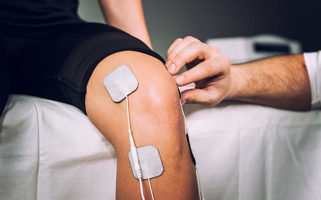
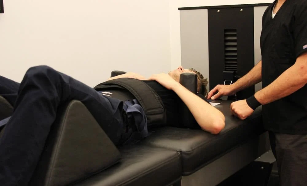
Figure 1: Patient undergoing treatment on the VAX-D Therapy Table
The blood supply to the nerve roots of the cauda equina is sensitive to compression. Even at pressures of only 5-10 mm Hg, the flow in over 20% of the venules was completely stopped (6). Flow in all the capillaries stopped at pressures between 20 and 50 mm Hg. A pressure of 30 mm Hg is slightly less than one pound per square inch, so solute transport is easily reduced. Even vertebral distractions (increased separation) of 1 or 2 mm per disc would reduce ligamental redundancy and help to restore canal/foraminal patency, reduce venous congestion and increase axoplasmic flow. Furthermore, the effects of lumbar spine lengthening may be sustained for a period of time after lumbar distraction has been stopped.
Twomey (7) placed lumbar vertebral columns removed from 23 male cadavers under 9 Kg of sustained traction for 30 min and measured an average increase in length of 9 mm. Thirty minutes after traction was removed, 13 of the 23 specimens had returned to baseline length, but the remaining 10 spines showed residual elongations ranging from 0.3 mm to 4 mm. Additionally, the data suggested that sustained traction had had a longer lasting effect on elderly spines. The mechanism of this residual deformation was not elaborated upon by the author, but disc rehydration may have been a factor since each column was soaked in normal saline and remained saturated by periodic additions of saline to a close fitting bag surrounding each column during the study.
That lumbar traction, if adequately applied, can effect physical change in patients suffering from back pain is well described by Gupta and Ramarao (8). They used water soluble contrast medium and epidurography to study 14 patients with prolapsed intervertebral disc syndrome before and after 10 to 15 days of continuous traction. Ten patients showed definite clinical improvement, with reduction in back pain and sciatica. Nine of these patients showed complete resolution of the defect on epidurogram and one of them showed partial reduction. The authors concluded that disc protrusion may be safely treated by traction. Mathews also demonstrated the effectiveness of lumbar traction in two patients by epidurography. Disc protrusions were decreased and an average vertebral distraction of 2 mm per disc space was shown in radiography (9). Judovich found that a traction force of approximately 26% of the body weight was needed just to overcome the resistance between the lower half of the patient and a (nonsplit) table (10).
Intuitively, lumbar traction should be successful in alleviating many of the conditions which cause low back pain and associated radiculopathy. Unfortunately, studies of clinical efficacy have yielded equivocal results. Previously, the successful application of lumbar traction has been limited by patient tolerance and the design of mechanical devices. Patients had difficulty tolerating the forces needed to relieve pain if delivered continuously. Furthermore, the thoracic corsets worn by patients to prevent movement on the table were uncomfortable, restrictred respiration, and can compromise venous return to the heart. Technological advances have now led to the development of equipment that has been found to achieve decompression of lumbar discs without stimulating the reactive reflexes of the lumbar musculature that can otherwise overcome efforts to effectively distract vertebral bodies.
The VAX-D therapy table is shown in Figure 1. The split table design eliminates frictional resistance between the patient and the table and allows controllable effective axial distraction tensions to be applied to the lumbar vertebral column. The equipment applies distractive forces in a gradual, progressive fashion, designed to achieve distraction of the vertebral bodies without eliciting reactive reflex muscular resistance. A portion of a typical chart recording of the tensile force applied to a patient's spine as a function of time is shown in Figure 2. Each decompression phase, during which the tension is increased, normally lasts for one minute. The force is increased more slowly in the latter part of the decompression phase. The tension is then gradually decreased, over a period of 30 sec, to about 20 pounds, which is maintained during the rest phase. Another cycle then starts. The avoidance of paravertebral muscle contraction, stimulated by homeostatic proprioceptor and axon reflex mechanisms allows the distraction of the vertebral bodies necessary to achieve decompression of the intervertebral disc. The therapy is administered via an automated logic control mechanism which systematically applies distractive tensions and rest periods in a cyclic fashion. The typical therapy session consists of 15 cycles of tension and relaxation. This periodic process allows patients to withstand stronger forces than can be tolerated when static techniques are used and it promotes accommodation and relaxation during the therapy session. The upper body is fixed by means of the patient grasping adjustable hand grips, designed to eliminate the use of a thoracic corset. Consequently, there is no risk of circulatory or respiratory compromise. The pelvis is secured with a specially designed harness that adjusts snugly and applies forces primarily to the lateral pelvic alae, thus minimizing anterior-posterior pressures and reactive muscle spasm during the distractive period of each cycle.
VAX-D treatment has been shown (11) to decompress the nucleus pulposus to pressures below - 100 mm Hg. This creates a tremendous potential diffusion gradient across the disc space, which is otherwise an avascular structure. Glucose and oxygen enter the disc at the end plate region while sulphate ions needed for the production of new glycosaminoglycans enter from the annulus fibrosis (12). Thus therapy may augment nutrient flow into the disc, facilitating structural restoration of the disc and promoting disc rehydration, since proteoglycans bind water (13). These effects may be cumulative with repetitive therapy sessions.
 Figure 2: chart recording of tension versus time for five cycles of the typical 15-cycle VAX-D Therapy session
MATERIALS AND METHODS
Data was collected from twenty two medical centers in the USA for patients who received VAXD therapy for low back pain. Only patients who received at least 10 treatments and had a diagnosis of herniated disc, degenerated disc, or facet syndrome, which was confirmed by imaging studies, were included in the study. The average number of treatments was 17 for facet syndrome, 19 for degenerative disc disease, and 20 for other diagnoses. The data contained the patients' assessment of their own pain, mobility, and ability to walk and sit. The pain scale ran from no pain (0) to severe pain (3). The mobility limitation scale was: No limitation (0), slightly limited (1), very limited (2), and completely immobile (3). The activity limitation scale was: walks frequently (0), walks occasionally (1), chairfast (2), and bedfast (3(). The treatment schedule, including the use of other modalities, the duration and frequency of VAX-D therapy, and medication was also recorded, as well as the patient's history. The symptoms were recorded at the beginning, mid-point, and end of the treatment schedule. The patients' satisfaction with the treatment was quantified as: not satisfied (0), slightly satisfied (1), very satisfied (2), and completely satisfied (3).
The data were divided into five groups:
- The first group which contained 34 cases, included all patients with extruded herniated discs, whether or not additional lesser problems were present.
- The second group contained 195 cases of multiple herniated discs, without extrusion, with or without degenerative disc disease.
- The third group consisted of 382 patients with a single herniated disc, regardless of degenerative disease.
- The fourth group contained 147 cases of degenerative disc disease, without herniation.
- The fifth group contained 19 cases with facet syndrome. Five cases of facet syndrome which had a pain reduction to 0 or 1 before 10 treatments, and one that had a reduction to 2, received less than 10 total reatments, so they were not included in the data base.
RESULTS
Table 2 shows how the average pain, mobility, and activity scores for the entire group of 778 patients improved during treatment. Although 51% of the pain reduction occurred during the first half of the course of treatment, 56% of the mobility improvement and 55% of the activity improvement occurred during the last half.
On a rating scale of 0 to 3, increases in spine mobility of one grade or more was seen in 77% of the patients with mobility limitations. Functional increases of 1 or more grades in the activity score was recorded in 78% of the patients who, before treatment were either unable to walk or capable of only limited walking. The coefficient of linear correlation14 between mobility and pain scores was 0.72. Between pain and activity the correlation was 0.60, and between activity and mobility it was 0.59. On a scale of 0 to 3, the average satisfaction with treatment was 2.4, which lies between 'very satisfied' and 'completely satisfied'.
In this study, 31 patients had previous lumbar disc surgery. MRI scans showed scar tissue that could potentially entrap nerve roots. Despite this, 84% of this group's pain scores and 71% of their mobility scores and 61% of their activity scores improved by one unit or more with therapy, and 65% of their pain scores were reduced to 0 or 1. Vertebral axial decompression was well tolerated. Vertebral axial decompression therapy outcomes: Earl
E. Gose et al.

| Table 2: Variation of average pain, mobility, and activity scores during treatment, and final outcome measures for the entire group | |||
|---|---|---|---|
| Pain (0-5 scale) | Mobility limitation (0-3 scale) | Activity limitation (0-3 scale) | |
| Before therapy | 4.10 | 1.81 | 1.24 |
| At midpoint | 2.62 | 1.30 | 0.80 |
| After therapy | 1.21 | 0.64 | 0.27 |
| Overall improvement | 71% | 65% | 78% |
| Improved by 1 unit or more | 92% | 77% | 63% |
DISCUSSION
We consider VAX-D therapy to be a primary treatment modality for low back pain associated with lumbar disc herniation at single or multiple levels, degenerative disc disease, facet arthropathy, and decreased spine mobility. Physiology (pain and mobility) and pathology correlate imprecisely. We believe that post-surgical patients with persistent pain or "Failed Back Syndrome' should not be considered candidates for further surgery until a reasonable trial of vertebral axial decompression has been tried.
Low back mobility increased subsequent to therapy and correlated well with pain reduction. Both of these factors are important in areas such as Workers Compensation and personal injury. Estimates of permanent partial impairment rely heavily on mobility aspects, as seen in the AMA Guides to the Evaluation of Permanent Impairment, 4th edition. Although allowance for pain is made in the percentage of impairment, the determination of impairment is made by determination of spine mobility using the range of motion model. By definition no patient can be assigned any impairment rating until maximum medical improvement (MMI) is reached. We submit that patients can usually be brought to a higher level of MMI by this therapy because of the anticipated improvements in mobility.
In summary, the pain, activity, and mobility scores were all greatly improved after therapy. VAX-D by its unique design may more precisely address the physiology of persistent low back pain than other conventional therapies. We consider it to be a front line treatment for degenerative spondylosis, facet syndrome, disc disease and nonsurgical lumbar radiculopathy.
REFERENCES
- Heldeman S. North America Spine Society: Failure of the pathology model to predict low back pain. Spine 1990; 15:718-724.
- Wheeler A.D. Diagnosis and management of low back pain and sciatica. Am Family Physician 1995; 52:1333-1341.
- Scientific approach to the assessment and management of activit-related spinal disorders. A monograph for clinicians. Report of the Quebec Task Force on spinal disorders. Spine 1987; 12(Suppl 7):1-59.
- Videman T, Saina S, Crites Battle M, Koskinen S, Gill K, Paanaman H. The long term effects of physical loading and exercise lifestyles on back-related symptoms, disability, and spinal pathology among men. Spine 1995; 20:699-709.
- Anderson GBJ, McNeill TW. Lumbar Spine Syndromes Evaluation and Treatment. New York: Springer-Veriag Wien, 1989: pp.1-215.
- Olmarker K, Rydeuik B, Holm S, et al. Effects of experimental graded compression on blood-flow in spinal nerve roots. J Orthop Res 1989; 7:817-823.
- Twomey LT. Sustained lumbar traction: An experimental study of long spine segments. Spine 1985; 10:146-149.
- Gupta RC, Romarao SV. Epidurography in reduction of lumbar disc prolapse by traction. Arch Phys MedRehabilitation 1978; 59:322-327.
- Mathews JA. Dynamic discography: A study of lumbar traction. Ann Phys Med 1968; IV:275-279.
- Judovich BC. Lumbar traction therapy-elimination of physical factors that prevent lumbar stretch. JAMA 1955; 159:549-550.
- Ramos G, Martin W. Effects of vertebral axial decompression on intradiscal pressure. J Neurosurg 1994; 81:350-353.
- Nachemson AL. The lumbar spine: An orthopaedic challenge. Spine 1975; 1:59-71.
- Ballard WT, Weinstein JN. Biochemistry of the intervertebral disc. In: Kirkaldy-Willis WH, Burton CV, eds. Managing Low Back Pain, New York: Churchill Livingston, 1992: pp.39-48.
- Gose EE, Johnsonbaugh R, Jost S. Pattern Recognition and Image Analysis, Upper Saddle River, NJ: Prentice-Hall PTR, 1996: pp.1-484.
If treatment success is defined as a reduction in pain to 0 or 1 on a 0 to 5 scale, the treatment was successful in 71% of the 778 cases. The success rate varied from 53% for the patients with extruded herniated discs, to 73% for patients with a single herniated disc. It was 72% for people with multiple herniated discs and 68% for facet syndrome. On a pain scale of 0 to 5, the people with extruded herniated discs had an average pain of 4.16 at the beginning of treatment and an average of 1.82 after treatment, a reduction of 56%. The cases of multiple herniated discs went from 4.13 to 1.18, a reduction of 71%. The patients with a single herniation had a reduction from 4.16 to 1.09, or 71%. The degenerative fisc cases reduced from 3.93 to 1.17, a 70% reduction. The patients with facet syndrome had a reduction of 4.00 to 1.13, a 72% reduction in pain. Overall, 71% of the patients experienced a reduction in pain to 0 or 1. The reduction in the average pain score was also 71%. One percent of the patients reported increased pain, 7% had no change, 92% improved by 1 unit or more, 87% improved by 2 units or more, and 70% improved by 3 units or more. A summary of these findings is shown in Table 1.
ABSTRACT
Low back pain is one of the most significant medical and socioeconomic problems in modern society. International guidelines call for evidence-based management for the pain and disability associated with musculoskeletal disorders. The purpose of this randomised controlled trial is to address the question of efficacy and appropriateness of VAX-D (Vertebral Axial Decompression) Therapy, a new technology that has been shown in clinical research to create negative intradiscal pressures, and has been shown to be effective in treating patients presenting with chronic low back pain (>3 months duration) with associated leg pain. Successful outcome was defined as a 50% reduction in pain utilising a 10cm Visual Analogue Pain Scale and an improvement in the level of functioning as measured by patient-nominated disability ratings. Patients were randomly assigned to VAX-D or to TENS which was used as a control treatment or placebo. The TENS treatment demonstrated a success rate of 0% while VAX-D demonstrated a success rate of 68.4% (P<0.001). A statistically significant reduction in pain and improvement in functional outcome was obtained in patients with chronic low back pain treated with VAX-D. (Neurol Res 2001; 23:780-784)
INTRODUCTION
Low back pain is a major cause of disability in today's society. According to the National Health and Medical Research Council (NHMRC), each year approximately 600,000 Australians present with low back pain as a recent illness. Although a high percentage of patients with acute low back pain recover within 4-6 weeks, a significant number of patients suffer from recurrences. Von Korff has studied the natural history and found that approximately 60% will have recurrences. (1) In a study of back pain in primary care, Von Korff and Saunders found that 60% to 75% improve in the first month, 33% report intermittent or persistent pain at year one, and 20% of patients describe substantial limitations at this time. (2) Klenerman et al demonstrated that 7.3% of individuals with acute low back pain who had not recovered by two months still reported high levels of pain and disability at twelve months after onset. (3) Chronic low back pain is increasing faster than any other disability, and 5-7% of the population will report their back problems as being a chronic illness. Fifty percent of work loss caused by back pain is accounted for by duration of disability for longer than 4 weeks. In Australia chronic low back pain affects more than 1,900,000 individuals and costs Australia more than 10 billion dollars each year.
International guidelines call for evidence-based management for the pain and disability associated with musculoskeletal disorders. Today's primary care practitioners have a comprehensive responsibility in the management of their patient's low back conditions, and they must be aware that recurrences after the presenting episode are likely. The literature suggests that for those who have not recovered by two months, management efforts should begin. (4)
Acute disc injury and discogenic pain is one of the primary processes leading to low back pain and lumbar radiculopathy, although the pathophysiologic mechanisms are still not well understood. It is believed that increases in disc pressures resulting from heavy lifting, vibrational and postural forces etc. are important factors in the pathogenesis of low back pain. The effects of disc hydraulics in herniations or protrusions may cause a mechanical deformation of the nerve roots and a compression-induced impairment of the vasculature. In addition, it has been found that the biochemical properties of the nucleus pulposus may induce a toxic or inflammatory reaction in the nerve root.
There have been many studies indicating that the disc and its associated pathology are identified as a primary cause of low back pain and lumbar radiculopathy. Hirsch stimulated various lumbar tissues in awake patients with the use of carefully placed needles. (5) Stimulation of the posterior portion of the annulus produced low back pain in many individuals. Furthermore, he was able to eliminate the pain by the injection of a minute volume of local anaesthetic into the annulus. Smythe and Wright placed nylon threads into various lumbar tissues while performing lumbar spinal operations. (6) During the postoperative period, they pulled on the threads and asked the patients to describe the location of any pain produced. The annulus fibrosus was the most common site of low back pain, and the compressed nerve root was responsible for sciatic pain. Tension placed on a normal nerve root resulted in no pain.
Falconer and associates published their observations made during exploration of the lumbar spine under local anaesthesia. (7) Murphy reported similar results in his small series of surgical cases. (8) Both authors concluded that the annulus and nerve root were the pain generating tissues. Wiberg in 1950, operating on 200 patients using local anaesthesia of the skin and muscles only, reported that pain emanated from the disc. (9) Kublisch operated on 193 patients using local anaesthesia and drew certain conclusions about the likely origin of back and leg pain. (10) Sciatica could only be produced by stimulation of a swollen, stretched, or compressed nerve root. Back pain was produced in the majority of cases by stimulating the outer layer of annulus fibrosus and the posterior longitudinal ligament.
If the disc is a major source of low back pain then applying specific target therapy for the treatment of disc pathology should improve patient outcomes. VAX-D is a primary, non-surgical treatment for the management of patients with disabling low-back pain and neurological symptoms associated with herniated and degenerative disc disease. Research has shown that the VAX-D table is a decompression device that is capable of reducing intradiscal pressures to negative levels. (11)
Successful reduction of intradiscal pressures with VAX-D represents a technological advance that should provide a means of addressing compressive disc pathology. Creating negative intradiscal pressure is likely to affect both the biomechanical and biochemical causes of discogenic pain. Patients suffering from discogenic pain and/or associated sciatic pain are seeking conservative treatment without the risks associated with injections and surgical procedures.
VAX-D incorporates advanced technology that permits the application of distractive tensions without eliciting reflex muscle guarding. Conventional traction devices have not demonstrated this ability or the ability to reduce intradiscal pressures to negative levels. Studies published in the medical literature report that intradiscal pressure either remains unchanged or increases during traction. (12) It has also been demonstrated that paraspinal muscles are not able to fully relax during conventional traction.
The beneficial effects of VAX-D decompression in the relief of peripheral nerve dysfunction has been previously reported in the literature, (13) and a multi-center outcome study reported that VAX-D treatment was successful in 71% of the 778 cases studied. (14)
This study was designed to evaluate the effect of VAX-D on chronic low back pain.
MATERIAL AND METHODS In association with Quintiles, the world's largest health care consultancy organisation for data analysis in clinical trials, a protocol was developed and then approved by the Human Research Ethics Committee at the University of Wollongong, New South Wales, Australia.
It was predetermined that the treatment would be considered a success if the patient attained a fifty percent (50%) decrease in pain, numerically on the Visual Analogue Scale (VAS). Absolute changes in pain score determined by VAS over time were analysed with repeated measures analysis of variance and t-test. In addition, improvements in disability were recorded on a patient nominated disability rating. Any level of improvement in disability was acceptable. The instruments for determination of these outcomes were supplied by the National Musculoskeletal Initiative of Australia. The study itself was to be conducted in the medical clinics of the VAX-D Spinal Institute and so to prevent bias in the data collection Quintiles were engaged to collect and analyse the data. TENS was selected as an appropriate placebo treatment as a means of establishing a plausible but (probably) ineffective control for an unblinded treatment.
Through advertisement in local papers forty-four patients with chronic low back pain greater than 3 months in duration, with associated leg pain, and a confirmed disc protrusion or herniation on CT Scan or MRI were selected and randomised into the two treatment methods, either VAX-D or TENS. The patients were randomised in sequential order and treatments were determined by a predefined central randomisation list.
The average duration of pain in the patient population was 7.3 years. The conditions for receiving either treatment including travelling to and from the clinic and duration of therapy were designed to be the same for both populations. Inclusion criteria for the study were: age 18-65 years; a minimum VAS score of 2; candidates must live within 45 minutes of the clinic location; capable of thoroughly understanding the information given and following protocol. All candidates signed an informed consent form.
Exclusion criteria were: osseous stenosis; unstable spine (bilateral pars defect or Spondylolisthesis of Grade II or greater); spinal surgical implants; shoulder problems which prevent compliance with VAXD therapy; spinal pain due to tumor, infection, or inflammatory disease; pregnancy; and previous VAXD therapy.
Patients randomised to VAX-D were treated according to the manufacturer's protocol. Patients lie on the split table device in a prone position. VAX-D utilises handgrips that the patient grasps with arms extended above the head to stabilise (restrain) the shoulder girdle and upper body. This is thought to be the most effective means of assuring that tensions applied to the pelvis are transmitted accurately along the linear axis of the spinal column during the procedure. The fact that the patient may release at any time during the treatment provides an important safety factor. A special harness designed to apply forces primarily to the lateral pelvic alae is fitted and tightened around the patient. The pelvic harness is connected to a tensionometer at the caudal end of the table. The function of the tensionometer is to provide constant feedback to the programmed logic control and operating system. During the VAX-D session a continuous chart recording is generated plotting the controlled time/energy progress of the entire procedure.
Tensions are applied to the lumbar spine in a cyclic fashion from the baseline tension up to the therapeutic range of fifty to ninety-five pounds. Each treatment session is thirty minutes in length and is comprised of fifteen cycles of decompression alternating with relaxation. Each decompression and relaxation phase may be individually varied as suitable for the particular treatment parameters.
A chart recorder prints the time energy curve for each decompression-relaxation cycle. This affords the technician a means of monitoring and adjusting the decompression process. Patients received VAX-D therapy five times per week for four weeks and then once per week for four weeks in accordance with protocol. All VAX-D treatments were administered by certified VAX-D technicians at four clinics in the Sydney area.
Patients randomised to TENS therapy received treatment at one of the four clinics. Electrodes were placed according to the manufacturer's protocol. Patients lay prone on a treatment table and received TENS for thirty minutes daily for twenty days then once a week for four weeks. All patients receiving TENS were monitored by a technician.
Neither group received any physical therapy modalities, epidural steroid injections or other treatments during the trial. Both patient groups were allowed to take non-narcotic pain relievers and antiinflammatory medication if necessary.
A 10-cm Visual Analogue Scale (VAS) for pain and a four-point disability rating scale were used to assess patient response. The level of pain on the VAS was recorded on a 10cm line marked at one end 'No Pain' and marked at the other end 'The Worst Pain Imaginable'. The written instruction to the patient was to 'please place a mark on the line below to indicate your current level of pain'. The self-nominated disability rating scale required patients to list the four activities that were most affected by their low back pain. These were scored according to the following criteria: 1 = cannot do at all; 2 = can do but severely limited; 3 = can do but slightly limited, 4 = can do without limitation.
Data was collected at the initiation of the study prior to randomization and at the end of the eight week treatment period in a separate interview. Success was defined as (equal to or greater than) a 50% improvement in the patient's pain and any improvement in their disability rating.
Patients were free to withdraw from the study on their own volition at anytime. The study treatment could be terminated prematurely if any of the following events occurred: patient wished to terminate his/her participation for whatever cause (two cases); the investigator judged it was in the best interest of the patient to withdraw (zero cases); the patient was unable to comply with protocol (zero cases).
The efficacy-evaluable population used for statistical analysis of efficacy is comprised of all patients who were randomised to study treatment, received at least 10 study treatments, had efficacy data recorded after Baseline, and satisfied the inclusion/exclusion criteria.
The primary efficacy measure in this study was the proportion of successfully treated patients in each of the treatment groups. The difference in proportions of successfully treated patients in each treatment group was tabulated and compared using Fisher's Exact Test and 95% confidence limits.
Successfully treated patients were to be followed up at six months to determine whether the successful outcome was sustained.
RESULTS
Forty-four patients were enrolled into the study. Twenty-two were randomised to each of the treatment groups. A summary of demographic characteristics for the 44 enrolled patients is presented in Table 1. Table 1: Demographic data
Characteristic Statistic All VAX-D TENS Two patients (4.5% of 44), Patient 029 and Patient 003, were regarded as having withdrawn/not completed the study according to the protocol. Patient 029, randomised to TENS, withdrew due to not wishing to continue and Patient 003, randomised to VAX-D, withdrew due to treatment no longer being required. No patients were withdrawn by the investigator. Patients 018 and 034 both randomised to VAX-D, did not comply with the study criteria and are therefore excluded from the efficacy-evaluable population. They both had a baseline VAS score less than 2 but this error of inclusion was not picked up until the completion of the trial. The efficacy-evaluable population therefore comprised of 40 patients: 19 patients randomised to VAX-D, 21 randomised to TENS.
| No of Patients | n | 44 | 22 | 22 | |
| Age (years) | Mean | 42 | 41 | 43 | |
| Range | 22 - 57 | 27 - 57 | 27 - 55 | ||
| Sex | Female | n | 21 | 11 | 10 |
| Male | n | 23 | 11 | 12 | |
| Race | White | n | 40 | 20 | 20 |
| Asian | n | 4 | 2 | 2 | |
| Chronicity | Mean | 7.3 | 8.4 | 6.2 | |
| Yrs of Pain | Range | 0.25 - 30 | 0.25 - 30 | 0.25 - 28 |
A summary of the data collected at baseline and post-treatment in the efficacy-evaluable population is presented in Table 2.
Table 2: Efficacy-evaluable population
| Characteristic | Statistic | VAX-D | TENS | |
|---|---|---|---|---|
| No of Patients | n | 19 | 21 | |
| Number of treatments | Mean | 24.1 | 18.0 | |
| Range | 18 - 36 | 10 - 24 | ||
| Baseline pain (VAS) | Mean | 5.99 | 5.44 | |
| Range | 2.1 - 8.7 | 2.7 - 8.5 | ||
| Post treatment pain (VAS) | Mean | 1.85 | 5.97 | |
| Range | 0 - 5.6 | 1.8 - 8.5 | ||
| Decrease in pain (%) | Mean | 69.1 | -17.1 | |
| Range | 11.1 - 100 | -123 - 33.3 | ||
| Disability | Pre-treatment | Mean | 2.2 | 2.2 |
| Rating | Range | 1.5 - 3 | 1.75 - 3.0 | |
| Post treatment | Mean | 2.9 | 2.2 | |
| Range | 2.0 - 4.0 | 1.5 - 3.0 | ||
| Improvement in disability rating | Mean | 33.8 | -2.23 | |
| (%) | Range | 0 - 100 | -36.4 - 50.0 | |
| Successful cases | n | 13 | 0 | |
| Percent | 68.4% | 0% | ||
In the efficacy-evaluable population the proportion of successfully treated patients was 13 out of 19 patients (68.4%) for the VAX-D treatment group compared to zero out of 21 (0%) for the TENS treatment group. There was a high statistically significant treatment group comparison p-value of <0.001. The 95% confidence interval for the difference in proportions of successfully treated patients, comparing VAX-D with TENS was 47.5% to 89.3%.
In the VAX-D group all patients recorded some improvement in their pain levels whereas in the TENS group 13/ 21 recorded an increase in pain.
At six-month follow-up, of the 13 successful cases, 2 have been lost to follow-up, 1 case suffered a significant other injury and of the remaining 10, seven have shown sustained success (ie. they still meet the criteria for successful outcome).
The results reported for the TENS group were less that that expected for a placebo control. The negative outcomes may have been due to the fact that the TENS patients (and the VAX-D patients) had to travel to and attend a medical clinic five days per week for four weeks, and one day per week for four weeks. This fact that both treatment groups had to travel to, and attend the clinic, was necessary to ensure that the only variable between the two groups was in the type of treatment that they received. The benefits of treatment in the VAX-D group clearly outweighed the negative effects of travelling, which became evident in the placebo group.
DISCUSSION
Disc stresses coupled with ongoing increased intradiscal pressures from mechanical loading may lead to failures in the normal biomechanics of the disc and progress to degeneration, posterior displacement of the nuclear material, annular disruptions and herniations. Other causative factors in the course of disc degeneration are negative diffusion gradients, reduction of the fluid content of the nucleus pulposus, and abnormal disc metabolism. With positive disc pressures throughout the day that are above diastolic pressure, the metabolism of the disc becomes anaerobic thus impeding the normal reparative healing abilities.
Proteolytic enzymes (matrix metalloproteinases) reside in the disc and have been implicated in disc degeneration. (15) The matrix metalloproteinases are regulated by specific inhibitors (TIMPS), cytokines (Interleukin-1) and growth factors. (16) Spinal loading may interfere with diffusion into the disc by reducing the gradient across the vertebral endplate. As disc metabolism becomes anaerobic, there is an accumulation of lactic acid, fall in pH, loss of chondrocyte and fibroblast function, and activation of the metalloproteinases.
Although the mechanism of action may not be fully understood the thixotrophic (17) properties of the nucleus material may facilitate nuclear migration toward the centre of the disc under negative pressures created by VAX-D.
It has been shown experimentally that elevated lactate levels and low pH in the disc prohibit disc proteoglycan synthesis and accelerates matrix degeneration (18).
Destruction of the proteoglycan matrix and fluid retention properties can lead to a degenerative cascade with loss of cellular reparative functions and vitality. The reduction of intradiscal pressures may enhance the diffusion gradient across the endplate into the avascular disc. It has been postulated that mechanisms that facilitate oxygen and nutrient uptake in the disc may exert a beneficial effect on the metabolism and restorative functions.
Successful reduction of intradiscal pressures with VAX-D therapy represents a technological advance in lumbar spinal treatment and is likely to affect both the biomechanical and biochemical causes of discogenic pain. The results from this study demonstrate that VAX-D is an effective treatment for the management of patients with chronic low back pain and is significantly superior when compared to TENS therapy. Analysis of the data demonstrated an attributable success rate of 68.4% for VAX-D. These findings are consistent with earlier studies by Gose E, Naguszewski W, Naguszewski R. (14)
The results of this prospective study demonstrated that VAX-D can achieve a statistically significant improvement in pain and functional outcome in managing patients suffering from disc related chronic low back pain.
ACKNOWLEDGMENTS
Australian National Musculoskeletal Initiative: For advice and instruction on the use of instruments for the outcome measures used in the present study.
Jane Ambrose, Biostatistician Quintiles: For statistical analysis of the data. DISCLOSURE Dr Russell Smart is contracted to and a shareholder in VAX-D Australasia Pty Ltd, a private company
that delivers VAX-D service in Australia.
REFERENCES
- Von Korff M. Studying the natural history of back pain. Spine 1994; 19(18 Suppl): 2041S2046S.
- Von Korff M., Saunders J. The course of back pain in primary care. Spine 1996; 21: 2833-2837.
- Klenerman L, Slade, PD Stanley IM. et al. The prediction of chronicity in patients with an acute attack of low back pain in general practice setting. Spine 1995; 20:478-484.
- Bogduk N, Evidence Based Clinical Guidelines for the Management of Acute Low Back Pain; The National Musculoskeletal Medicine Initiative NHMRC;Nov 1999
- Hirsch C. An attempt to diagnose the level of disc lesion clinically by disc puncture. Acta Orthop Scand 1948;18:132-140
- Smythe MJ. and Wright V. Sciatica and the intervertebral disc. An experimental study. J Bone Joint Surg (Am) 1958;40:1401-1418
- Falconer MA; McGeorge M, Begg AC; Observations on the cause and mechanism of symptom production in sciatica and low back pain. J Neuro Neurosurg Psychiatry 1948;11:13-26
- Murphy F. Experience with lumbar disc. Clin Nerurosurg 1973;20:1-8
- Wiberg G. Back pain in relation to the nerve supply of the intervertebral disc. Acta Orthop Scand 1950;19:211-221
- Kublisch S, Ulstrom C, Michael C. The Tissue of Low Back Pain and Sciatica: A Report of Pain Response to Tissue Stimulation During Operations on the Lumbar Spine Using Local Anesthesia. Orth Clinics of North Am 1991; 22:181-187
- Ramos G, and Martin W. Effects of vertebral axial decompression on intradiscal pressure. J. Neurosurg 1994; 81: 350-353.
- Anderson G, Schultz A, Nachemson A. L. Intervertebral Disc Pressures During Traction. Scand J Rehabil Supp 1983; 9:88-91
- Tilaro F, Miskovich D. The Effects of Vertebral Axial Decompression On Nerve Sensory Dysfunction; Can J of Clinical Medicine 1999 Vol 6 No 1: 2-7
- Gose E, Naguszewski W, Naguszewski R. Vertebral axial decompression therapy for pain associated with herniated or degenerated discs or facet syndrome: An Outcome Study. J Neurological Research 1998 Vol 20:186-190.
- Bogduk N. Clinical Anatomy of the Lumbar Spine and Sacrum. Third Edition. Churchill Livingstone 1997.
- Fujita K, Nakagawa T, Hirabayashi K, Nagai Y. Neutral Proteinases in Human Intervertebral Disc. Role in Degeneration and Probable Origin. Spine 1993; 18:1766-1773
- Nachemson A. Elfstrom G. Intravital Dynamic Pressure Measurements in Lumbar Discs. Scand
J. Rehabil Med (Supp) 1970; 1:4-40
18. Matsui Y, Macda M, Nakagami W, Iwata H. The Involvement of Matrrix Metalloproteinases and Inflammation in Lumbar Disc Herniation. Spine 1998; 23:863-69
An Overview of Vertebral Axial Decompression
Dr. Frank Tilaro, M.D Canadian Journal of Clinical Medicine Vol 5, No 1, January 1998
INTRODUCTION
Low back pain is a growing epidemic among industrialized societies. In the United States it is the most common work related disorder. The cost to industry is staggering, with estimates running 20 billion dollars or more annually (4,42). Total payments for a single Workman's Compensation claim may be as high as $100,000. Abenhain and Suissa studied the 1-year incidence of work related low back pain in the province of Quebec for the year 1981 (1). Work absence due to back pain has an incidence of 1.4%. Seventy-four percent of work related injuries return to work within 1 month. 7.4% were out of work for more than 6 months. 75% of the direct total cost was borne by 10% of the absentees. Recurrence rates were 20% at 1 year and 36% after 3 years.
Men had higher recurrence rates than women; drivers and nurses had higher recurrence rates than other occupations. The recovery rate of the Quebec workers is similar to other countries. After 1 year 4.3% remained absent from work. Incidence rates of compensated back injuries by industrial sector showed that foresters and miners are at the top with 4.9% and 3.3% respectively.
BACK PAIN - A DIAGNOSTIC AND THERAPEUTIC DILEMMA
Effective diagnosis and therapy requires thorough knowledge of spinal biomechanics. Our approach to back pain has been centered on a patho-anatomical model but unfortunately the model frequently fails to comply with the clinical picture. The Quebec Task Force Report stated: "There is so much variability in making a diagnosis that this initial step (i.e. clinical assessment) routinely introduces inaccuracies which are then further confounded with each succeeding step in care ."(43) Adding to the confusion is the belief by too many physicians, patients and insurers that high tech imaging is the standard for establishing a diagnosis. However, the high rates of false positive and false negative findings point to the inadequacies of these studies in identifying the pain generating lesions (8,19,20,48,49). Nachemson states: "A confirmatory imaging study is indicated only if surgery is contemplated. Clinical symptoms and findings remain the most important basis for diagnosis."(28)
The natural history of low back pain with and without radiculopathy has been described (10,37,47). Spontaneous regression takes place in 80% to 90% of patients with low back pain by 6 weeks and a significant percentage of patients with sciatica report a satisfactory response to conservative medical management. Studies on disc surgery emphasize inappropriate patient selection as the cause for surgical failure (11,16,30,44). In Kramer's address to the International Spine Society he emphasized that the surgical failed back syndrome is the worst possible scenario a spine surgeon faces (22). In North America the incidence for this iatrogenic disease is about 15%, compared to 5% with most European countries (28). Comparisons between the United States and Europe indicate that the frequency of surgery in the U.S. is four times greater (11). Statistics from the Back Pain Outcome Assessment Team compiled from 1979 to 1987 indicate a rapidly growing number of disc excision and fusion operations performed each year, further escalating the cost (11,44).
Studies of the various surgical procedures largely lack validity and controlled prospective studies are rare (7). A randomized study by Revel demonstrated percutaneous discectomy has little value (32) and the same is true for laser discectomy. Chemonucleolysis is superior to saline injection but inferior to surgical discectomy. While chemonucleolysis had its followers for a period of time, it has fallen into disrepute because of the serious side effects including anaphylaxis and myelitis and should no longer be considered an option. There are not any studies demonstrating the superiority of one particular surgical intervention and there is no support for adding a fusion to a routine discectomy (11,27,28).
THE VAX-D THERAPEUTIC TABLE
The VAX-D therapeutic table (Vertebral Axial Decompression) addresses the functional and mechanical aspects of discogenic pain and disease. The table was invented by Dr. Allan Dyer, former Deputy Minister of Health from Ontario and a pioneer in the development of the external cardiac defibrillator. The table is designed to apply distraction tension to the patients lumbar spine without eliciting reflex paravertebral muscle contractions. The patient lies in a prone position, the upper body is over the stationary portion of the table, and the body is restrained by the patient holding on to adjustable handgrips which can be released at anytime for safety.
The table is a split table design, whereby distraction tensions are applied to the patient through a pelvic harness attached to a tensionometer and by separation of the movable part of the table. The distraction-relaxation cycles are automated or variably timed. Distraction tensions and rates are continuously monitored and measured by the tensionometer and the output is shown on a digital gauge and captured on a pen-write printout. The table exerts its effects through decompression of the intervertebral discs.
Dr.'s G. Ramos and W. Martin of the Departments of Neurosurgery and Radiology at the HCA Rio Grande Regional Hospital, McAllen, Texas studied intradiscal pressure during VAX-D therapy.(30) The patient population was comprised of individuals with unresolved low back pain who were referred for neurosurgical consultation. Previous management programs included conventional bedrest, medications, physical therapy, and or chiropractic treatments. Depending on the diagnosis and findings of the examinations, patients were assigned to one of the following study groups: Intradiscal Pressure Study and Clinical Outcome Assessment Study. Patients with a subligamentous herniation at L4-5 who were candidates for percutaneous discectomy were included in a study of intradiscal pressure manometry. The pressure measurements were recorded by two different methods; an Ohmeda pressure transducer connected to a Hewlett Packard pressure monitor via a saline bridge and a Camino fiberoptic intracranial transducer adapted for intradiscal measurements. Both transducers were recalibrated after each procedure utilizing a Pneumatic Calibration.
ANALYZER
The transducers were placed in the L4-5 disc under A-P and lateral fluoroscopy. With the catheter in place the patient was placed prone on the VAX-D table. Various decompression tensions from 50 to 100 pounds were applied. The distraction tensions and the resulting changes in intradiscal pressure were observed on digital readout and recorded on a graph tracing produced by the chart recorder. Intradiscal pressures were significantly reduced to minus 150-160mmHg. It was observed that a threshold distraction tension was necessary to develop negative pressures in the disc. The extent of decompression measured in mmHg follows an inverse relationship to the tensions applied.
The significance of this study cannot be overemphasized. The reduction of intradiscal pressure to negative levels has far reaching therapeutic implications. Prior to the introduction of VAX-D, a non surgical method for disc decompression was unavailable. In numerous studies, conventional traction has never demonstrated a reduction of intradiscal pressure to negative ranges, on the contrary many traction devices actually increased intradiscal pressure most likely secondary to reflex muscle spasm .(5)
INDICATIONS AND GENERAL USE OF THE VAX-D THERAPY TABLE
VAX-D is indicated for patients with low back pain that has been unresponsive to conventional therapy for 6-8 weeks. Patients with radiculopathies are also candidates. The presence of a neurological deficit does not affect patient eligibility since studies have revealed the outcome in patients with neurological deficits was not affected by surgical or medical management.(15) The presence of a rapidly progressive neurological deficit is an indication for surgery. Patients presenting with a fusion and the post surgical failed back syndrome may also be candidates.
Contraindications for VAX-D therapy include infection, neoplasm, osteoporosis, bilateral pars defect or Grade 2 spondylolysthesis if unstable, fractures, the presence of surgical hardware in the spine, and the cauda equina syndrome. Patients with lateral stenosis and central stenosis may respond if severe secondary changes are not present in the vertebra. The patient should be evaluated by a therapist or physician prior to initiating therapy and routine spine films are necessary to rule out any contraindications. A CT scan or MRI is not necessarily a prerequisite before therapy. The daily therapy sessions are administered by a trained VAX-D technician. All VAX-D technicians are encouraged to complete a certification exam. Treatments are administered on a daily basis for approximately twenty sessions and are routinely given Monday through Friday. An occasional patient may require a short maintenance period where 2 to 3 treatments a week are given for 2 to 4 weeks post therapy. The average patient has required 20-25 sessions. Each session is comprised of 15 cycles, each cycle being 1 minute in distraction and 1 minute in relaxation.
The table is designed to be operator friendly. With the patient standing, a specially made pelvic harness is fitted and tightened on the patient. The patient lies prone on the table with the lower portion of the belt placed at the level of the table separation point. The adjustable handgrips are positioned such that the elbows remain straight. Repositioning and tightening of the pelvic harness is completed at this point. The harness is attached to a movable pretension housing that maintains a baseline tension of 20 lbs. throughout the rest phase. Once the pretension is set the treatment cycles may begin. The Ramos study indicated that 50 lbs. of tension was the threshold tension necessary to develop negative intradiscal pressures. The p.s.i. is slowly increased until tensions of 60-80 lbs. are developed, this may take 3 to 4 days of therapy. Some patients have required 90-100 lbs. of tension for a full therapeutic effect.
Pain distribution frequently changes during or immediately after therapy. A phenomenon called centralization first observed by McKenzie (24) has been noticed during a course of VAX-D therapy. Centralization is the process by which the pain pattern migrates from a peripheral distribution to a more central or proximal location and is an indication of a favorable clinical outcome. Centralization of pain patterns may be associated with increased central back pain, but this should be interpreted as a positive sign and is likely secondary to stretching of the posterior longitudinal ligament as the lateral distortion of the disc retracts to a more concentric position. Centralization is a predictable prognostic indicator for symptomatic discs and annular competence (12) The observed occurrence of centralization during VAXD therapy in a patient who initially could not centralize their pain pattern implies healing of the annulus as a result of VAX-D therapy.
As higher distraction tensions are reached few patients may report an increase in pain of a different quality. Overstretching of the soft tissues in the back likely represents the cause of this pain and the patient should be treated by decreased distraction tensions, so as not to traumatize the soft tissues. The development of a sharp, burning, radiating pain during therapy could represent the stretching of an entrapped nerve. Since the breakdown of scar tissue is an objective, the patient should continue but distraction tensions should be reduced such that any pain elicited does not last more than 15-20 minutes post therapy. Distraction forces are then slowly increased over the ensuing days.
No serious side effects have been reported with VAX-D therapy. A limiting factor affecting the patient's tolerance to therapy is stress to the shoulder girdle and rotator cuff. This may be mitigated by placing a roll under the axilla of the affected side. Should a patient have discomfort from any cause, they may release the handgrips at any time. This adds an important safety factor to the treatment.
MECHANISM OF ACTION
An understanding of spinal biomechanics is necessary to appreciate VAX-D's mechanism of action, to effectively treat and diagnose spinal disorders, and to objectively review old and new therapies. The literature is replete with biomechanical data. Vogel and Stahl have carried out in vitro experiments on intradiscal movements with symmetrical and asymmetrical loading. (21) With symmetrical loading, the nucleus expands and is retained by the annulus. By contrast, if the disc is subjected to an asymmetrical load, the nucleus migrates to the area of least load or resistance.(38) With removal of the load, the nucleus moves from an eccentric to a more concentric position within the disc. Relocation can be accelerated by compression in the opposite direction or by distraction. (21) The annulus of a normal disc can restrain the nuclear movement, but when the elastic properties of the annulus are compromised the structures become susceptible to injury. Fissures and ruptures develop which allow the nucleus to migrate.
Fissures are normally present by 30-35 years of age and increase with advancing age. Fragment sequestra appear as a result of age and trauma. These fragments can move independently and result in protrusions and disc prolapses. Migration of nuclear material and sequestra is influenced by compressive forces, shearing, and increased intradiscal pressure. (24)
Epidemiological data and scientific data have demonstrated that prolonged or repetitive flexion loads stress the posterior annulus resulting in discogenic pain and in some patients disc herniation.(3,25) Adams and Hutton carried out experiments with gradual loading of the disc and concluded that disc prolapses can occur with a sustained flexion load.(2) Hickey and Hukins performed experiments with bending and torsion and demonstrated that the annulus failed posteriorly.(17) Shirtzi-Adl demonstrated that disc fiber layers are most loaded in flexion and least in extension.(40) Nachemson's research on intradiscal pressures showed pressures were highest with flexion.(26) The outer third of the annulus is innervated by the sinuvertebral nerve. Any asymmetrical load associated with elevated intradiscal pressure can result in overstretching and fatigue of the annulus, thereby stimulating the mechanoreceptors in the outer third of the annular wall. Eventually, fissure's will develop in the annulus which can lead to herniation of the central mass of the nucleus. By reducing intradiscal pressure with VAX-D therapy, a therapeutic and prophylactic effect can be realized.
Numerous studies utilizing discography have helped us to understand the role of the disc as a pain generator. Provocational discography is the standard test for discogenic pain.(41) Its reliability has been questioned and opponents generally refer to the work of Holt, but his study has been refuted on methodological grounds.(9,18,41) Recently a pathological marker of symptomatic disc disruption called the high intensity zone (HIZ) was demonstrated on MRI using spin echo gradient heavy T2 imaging.(6,38) An HIZ is evident in the posterolateral view on the sagittal section, which on provocation discography corresponded to a Grade 3 radial tear. The high signal intensity represents fluid within the fissure that may be causing pain either by chemical irritation or mechanical traction of the sinuvertebral nerve. By its cyclic action and ability to reduce intra-negative pressure, VAX-D therapy could displace the fluid to the internal portion of the nucleus thereby ameliorating pain and enhancing healing of the annulus.
Donelson demonstrated it is possible to predict annular competence with the McKenzie mechanical assessment protocol.(12) In his study patients were separated into centralizer's and non centralizer's. Discography was performed in both groups. Centralizer's tended to have an intact annulus or Grade 1-2 tears. Non centralizer's had a disrupted annulus , that is fissures to the outer third of the annular wall or Grade 3 tear. This is very exciting news for those who appreciate the centralization phenomenon because it allows us to clinically assess the competency of the annulus. Patients who centralize on initial evaluation may be treated with specific exercise. Patients who do not centralize on initial examination are excellent candidates for VAX-D therapy. With such an approach, the patients disposition regarding effective therapy is known immediately, which arguably translates to reduced disability and reduced cost.
VAX-D therapy has been shown to convert non centralizer's to centralizer's during or after successful VAX-D therapy. This implies VAX-D is conducive to annular healing. Asymmetrical loading of the disc and increased intradiscal pressure is partly responsible for internal derangement, disc degeneration and herniation. Changes in intradiscal pressure also play a prominent role in affecting nourishment of the disc since the disc is an avascular structure and receives its nourishment primarily by diffusion. Intradiscal pressure that is greater than capillary pressure in the vertebral body impedes oxygen diffusion to the disc which in turn impedes healing.(13) Reducing intradiscal pressure with VAX-D creates a diffusion gradient into the disc allowing nourishment to proceed. Solutes such as oxygen have a steep concentration gradient across the disc, with the peripheral concentration 20-30 times more than the concentration at the center of the nucleus. The availability of oxygen may be inadequate to meet the metabolic requirements required to heal a damaged annulus, and higher concentrations of lactate have been measured within the central portion of the disc. By reducing intradiscal pressure, VAX-D therapy creates a diffusion gradient thereby enhancing solute transfer. High levels of lactate could facilitate chondrocyte cell death as well as increase the activity of degradative enzymes further promoting the loss of the proteoglycan cell matrix. A vicious cycle is produced, accelerating disc degeneration. The mechanical effects of fluid loss during a compressive load are followed by a slow rate of disc deformation termed creep. The rate of creep is faster in a damaged or degenerated disc than a normal disc. Both vibration (overstress) and inactivity (understress) affect the rate of creep and disc degeneration. Pigs who were subjected to vibratory creep had lower levels of oxygen and sulfate transport, and higher levels of lactate within the disc.(13)
Somewhere between the overstress of vibration and the understress resulting from inactivity is an optimum mechanical environment.
Through its action, VAX-D may be capable of restoring that environment, enhancing healing of the disc and retarding degeneration. The pathophysiology of nerve root compression has been described by Rydevik.(33,34,35) The nerve root ganglion has an extensive venous plexus, which if obstructed, results in venous hypertension and endoneural edema, leading to hypoxia, ischemia and pain. External decompression with VAX-D therapy can be expected to relieve venous hypertension and reverse the pathognomonic process.
By significantly reducing intradiscal pressure, VAX-D promotes retraction of the herniation into the disc. VAX-D therapy could possibly shear a herniation from its connection to the central nucleus, creating a severed fragment within the spinal canal. This sequestered disc is susceptible to small vessel invasion and digestion as a result of contact with the epidural space. Reinforcing this theme is the study by Modic et al who studied the natural history of disc herniation by MRI in patients with acute radiculopathy and discovered that large (6mm) sequestered hernias were the first to undergo spontaneous resolution.(23)
I have studied sensory nerve dysfunction measured before and after VAX-D therapy, in order to determine the effect of VAX-D on nerve root compression. The results from this study are very significant and the data will be published in the future. Inflammation very likely plays a role in disc pathology and herniation, but the response to anti-inflammatories is rather disappointing. Saal found high levels of Phospholipase A2 in human disc samples removed at surgery in patients with radiculopathy.(36) As the enzyme responsible for liberation of arachidonic acid from cell membranes, Phospholipase A2 is the rate limiting step in the production of prostaglandins and leukotrienes. Controlled studies have shown that anti-inflammatories are not useful for acute sciatica,(46) but since solute transfer to the disc may be enhanced by VAX-D therapy, the administration of antiinflammatories during VAX-D therapy may result in higher concentrations of the drug within the disc, neutralizing inflammatory mediators responsible for nerve root inflammation and some forms of discogenic pain. As previously mentioned, the fluid within the HIZ is thought to be an inflammatory fluid and VAX-D may be able to effectively pump this fluid into the central nucleus where it is not possible for it to exert an inflammatory effect. The fissure may be able to approximate its borders and heal once the fluid is pumped out.
VAX-D VS. TRACTION
The VAX-D table is an external decompression device and this separates it from conventional traction. Studies verifying decompression of the disc and nerve root are now available for VAX-D. I reviewed the literature and I could not find any available data that conventional traction reduced intradiscal pressure to the negative range nor are there any studies on conventional traction showing beneficial effects in nerve root compression and conditions associated with discogenic dysfunction.
Many patients who receive VAX-D therapy have had chronic back pain and failed numerous modalities including traction. Their positive response to VAX-D therapy, after having failed conventional therapy and traction confirms VAX-D's assertion that it is not conventional traction.
CLINICAL STUDIES
The Acute Low Back Distress Study was conducted by the John P. Robarts Research Institute, London Ontario. The efficacy of VAX-D therapy was established with this study. The parameters measured were severity and duration of pain and disability, including analgesic requirements, and the presence and degree of neurological involve-ment. One hundred and ten patients were entered into the study.
The treatment was considered a success if the baseline aggregate score for pain and disability was reduced by 50% after 10 treatments of VAX-D therapy. Sixty-six percent of the patients achieved success according to the study protocol. Prior to therapy the aggregate score for pain and disability was
5.1 and after 10 treatment sessions in the successful group it was 1.2.
The Clinical Outcome Assessment Study was conducted at McAllen HCA Hospital by Dr. G Ramos.(31) Fifty-two patients completed VAX-D therapy as the primary modality. Thirty-eight patients (73%) achieved a positive outcome with remission of their low back pain symptoms and a return to functional levels of activity. Ninety percent of the recovered group were suffering from disc herniations, the majority (89%) being subligamentous while 11% had extruded herniations. Neurological deficits did not compromise the response to therapy.
Review of the patients clinical findings for those who achieved remission showed that 33% exhibited neurological deficits and 73% had sciatic pain prior to therapy with VAX-D. Dr. E. Gose, Dr. W Naguszewski, and Dr. R Naguszewski have completed an outcome study of VAX-D therapy from over twenty medical centers that included over 700 patients. Patients with back pain, with or without leg pain were included in the study as well as the failed surgical back patient. All patients had a diagnosis of a herniated disc, degenerative disc or facet syndrome. The authors are very enthusiastic about the outcome and have prepared a detailed report of their findings which have been accepted for publication in another respected medical journal.
An outcome study at Columbia Hospital, Tulsa OK. is currently being conducted that shows a level of success consistent with the above study.
SUMMARY
VAX-D therapy addresses the biomechanical aspects of discogenic disease and achieves its objective through decompression. It should be utilized in patients with low back pain, with or without radiculopathy who have failed conventional therapy (physiotherapy and chiropractic), and should be utilized prior to addressing surgery. By addressing the altered biomechanics responsible for disc disease, the VAX-D therapeutic table not only alleviates pain but has been shown to exert a beneficial effect on a major determinant in the equation responsible for discogenic disease, that is elevated intradiscal pressure.


Further analysis of future and unpublished research should be considered to further validate the therapeutic benefit of VAX-D therapy, however, these clinical studies have shown it to be effective in back pain syndromes with or without radiculopathy including herniated discs and internal disc disruption.
The chronic back pain patients and surgical patients are very costly to society. Since many of these patients are responsive to VAX-D therapy, this unique non-obtrusive means of managing the common forms of debilitating low back pain associated with discogenic disease could represent a considerable savings.
ABOUT THE AUTHOR
Dr. Frank Tilaro is a board certified internist who has been practicing orthopaedic medicine for the last 12 years. Dr. Tilaro has restricted his practice to orthopaedic medicine. He is a member of the American Back Society, the McKenzie Institute, the American Association of Orthopaedic Medicine and the British Institute of Musculoskeletal Medicine. Dr. Tilaro is medical director for The Advanced Spinal Institute in Ogden Utah and director of education and clinical research for VAX-D Medical Technologies, Palm Harbor, Florida. Dr. Tilaro resides in Ogden, Utah and can be reached at 1-801-698-0270 begin_of_the_skype_highlighting 1-801-698-0270 end_of_the_skype_highlighting.
REFERENCES
- Abenhain L, Suisse S, Rossignol M. Risk of recurrence of occupational back pain over three year follow-up. British J Ind Med 1988; 45:829-33.
- Adams A, Hutton WC. Gradual disc prolapse. Spine 1985; 10:524-31.
- Adams M, Green T, Dolan P. The strength in anterior bending of lumbar intervertebral discs. Spine 1994; 19:2197-2203
- Akeson WH, Murphy RW. Low back pain. Clin Ortho 1977; 129:2-3.
- Andersson GB, Schults AS, Nachemson AL. Intervertebral disc pressures during traction. Scand J Rehabil Med 1983; 9:88-91
- Aprille C, Bogduk N. High intensity zone: a diagnostic sign of painful lumbar discs on magnetic resonance imaging. The British Journal of Radiology 1992; 65:361-369.
- Bloch R, 7. Bloch R, Davis DO, Dina TS. Methodology in clinical back pain trials. Spine 1987; 12(5): 430-08.
- Boden SD, Patronas NJ, Weisel SW. Abnormal magnetic resonance scans of the lumbar spine in asymptomatic subjects: a prospective investigation. J Bone Joint Surg {Am} 1990. 72:403-08.
- Bogduk N, Modic MT. Controversy: lumbar discography. Spine 1996.21:402-04.
- Bush K, Cown N, Katz DE, Gishen P. The natural history of sciatica associated with disc pathology. A prospective study with clinical and independent radiologic follow-up. Spine 1992; 17(10):1205-12.
- Deyo RA, Cherkin DC, Loeser JD, Bigos SJ, Ciol MA. Morbidity and mortality in association with operations on the lumbar spine, The influence of age, diagnosis, and procedure. J Bone Joint Surg {AM} 1992; 74 (4):536-43.
- Donelson R, Aprille C, Medcalf R, Grant W. A prospective study of centralization of lumbar and referred pain. A predictor of symptomatic discs and annular competence. Spine 1997; 22(10):1115-1122.
- Frymoyer JW. The adult spine. Principles and practice. Raven Press, New York 1991.
- Gray ICM, Main CJ, Waddell G. Psychological assessment in general orthopaedic practice. Clin Orthop 1985; 194:258-63
- Hakelius A. Prognosis in sciatica. Acta Orthop Scand (suppl) 1970. 129:1-76
- Herron LD, Turner T. Patient selection for lumbar laminectomy and discectomy with a revised objective rating.Clin Orthop 185; 199:145-52.
- Hickey D, Hukins D. Relation between the structure of the annulus fibrosis and the function and failure of the intervertebral disc. Spine 1985; 5:106-114
- Holt EP. The question of lumbar discography. J Bone Joint Surg 1968; 50A:720-5.
- Jensen MC, Brant-Zawadski, Obuchowski N, Modic MT, Malksalan D, Ross JS. Magnetic resonance imaging of the lumbar spine in people without back pain. N Engl JMed 1994.331:69-
73.
- Komberg M. Discography and magnetic resonance imaging in the diagnosis of lumbar disc disruption. Spine 1988; 14:908-18.
- Kramer J. Intervertebral disk diseases. Causes, diagnosis, treatment and prophylaxis. George Theime Verlag Stuttgart, New York, 1990.
- Kramer J. Presidential address: natural course and prognosis of intervertebral disc diseases. Spine 1995; 20(6):635-39.
- Modic M, Ross J, Obuchowski N, Browing K, Cianflocco AJ, Mazanac D. Contrast enhanced magnetic resonance imaging in acute lumbar radiculopathy: a pilot study of the natural history. Radiology 1995. 195(2):429-35.
- McKenzie RA. The lumbar spine: Mechanical diagnosis and therapy. Waikane, New Zealand: Spinal Publications, 1981.
- McNally DS, Adams MA, Goodship AE. Can intervertebral disc prolapse be predicted by disc mechanics.Spine 1993. 18:1525-1530
- Nachemson AL. The influence of spinal movements on the lumbar intradiscal pressure and on the tensile stresses in the annulus fibrosis. Acta Ortho Scand. 1963; 33:183.
- Nachemson AL. Fusion for low back pain and sciatica. Acta Orthop Scand 1985; 56(4):285-86.
- Nachemson AL. Lumbar disc herniation-conclusions. Acta Orthop Scand (Suppl 251)1993;64.
- North RB, Campbell JN, James CS, Conover-Walker, Wang H, Paintadosi S, Rybock JD, Long DM. Failed back surgery syndrome: 5-year follow up in 102 patients undergoing repeated operation. Neurosurgery 1991; 28(5):685-91.
- Ramos G, Martin W. Effects of vertebral axial decompression on intradiscal pressure, JNeurosurg 1994; 81:350-353.
- Ramos G. Personal communication.
- Revel M Payan C, Vallee C, Laredo JD, Lassae B, Roux C, Carter Salomon C. Delmas E, Roucoules J, Beauvais C, Savy JM Chiecheportiche V, Bourgeois P, Smadja M, Hercot O, Wybier M, Cagan GM, Blum-Boisgard C, Fermanian J. Automated percutaneous discectomy versus chemonucleolysis in the treatment of sciatica. A randomized multicenter trial. Spine 1993; 18(1):1-7.
- Rydevik B, Brown MD, Llundborg G. Pathoanatomy and pathophysiology of nerve root compression. Spine 1984; 9:7-15.
- Rydevik B, Myers R, Powell HC. Pressure increase in dorsal root ganglion following mechanical compression.Closed compartment syndrome in nerve roots. Spine 1989; 14:574-76.
- Rydevik B, Holms S, Brown NM. Nutrition of spinal nerve root. Trans Orthop Res Soc 1984; 9:276.
- Saal JS, Franson R, Dobrow R, Saal JA, White A, Goldwaite N. High levels of inflammatory phospholipase A2 activity in lumbar disc herniations. Spine 1990; 15(7):674-78.
- Saal JA, Saal JS, Herzog R. The natural history of lumbar intervertebral disc extrusions treated non operatively.Spine 1990; 15(7):683-86.
- Schellhas K, Pollei SS, Gundry C, Heithoff K, Lumbar disc high-intensity zone. Correlation of magnetic resonance imaging and discography. Spine 1996; 21:79-86.
- Schnebel B. Simmons J, Chowing J, Davidson R. A digitizing technique for the study of movements of intradiscal dye in response to flexion and extension of the lumbar spine. Spine 1988; 13:309-12.
- Shiratzi-Adl A. Biomechanics of the lumbar spine in sagittal /lateral moments. Spine 1994; 19:2407-12.
- Simmons JW, Aprille CN, Dwyer AP, Brodsky AE. A reassessment of Holt's data on the "question of discography." Clin Orthop 1988; 237:120-4.
- Spengler D, Bigos S, Martin N, Seh J, Fisher L, Nachemson A. Back injuries in industry: A retrospective study.Overview and cost analysis. Spine 1986; 11:241-45.
- Spitzer WO, Leblanc FE, Dupuis M. Scientific approach to the assessment and management of activity-related spinal disorders. Spine 1987; 12:7S.
- Turner JA, Ersek M, Herron L, Deyo R. Surgery for lumbar spinal stenosis, attempted meta-analysis of the literature. Spine 1992; 17(1): 1-8.
- Vanharanta H, Sachs BJ, Spivey MA, Guyer RD, Hochshuler SH, Rashbaum RF, Johnson RG, Ohnmeiss D, Mooney V. The relationship of pain provocation to lumbar disc deterioration as seen by CT/discography. Spine 1987; 12(3):295-98.
- Weber H, Holme I, Amlie E. The natural course of acute sciatica, With nerve root symptoms in a double blind placebo controlled trial evaluating the effect of piroxicam (NSAID). Spine 1993; 18:1433-8.
- Weber H. Spine update. The natural history of disc herniation and the influence of intervention. Spine 1994. 19(19):2234-38.
- Weisel SW, Tsourmas, Fefer HL, Citrin CM, Patronas N. A study of computer assisted tomography: The incidence of positive CAT scans in an asymptomatic population. Spine 9:549-
51.
49. Zuckerman J, Derby R, Hsu K et al. Normal magnetic imaging with abnormal discography. Spine 1988; 13:1355-9.
Efficacy of VAX-D on chronic low back pain: Study of Dosage Regimen
Gustavo Ramos M.D, Department of Neurosurgery and Radiology, Rio Grande Regional Hospital, McAllen Texas
ABSTRACT
Vertebral Axial Decompression (VAX-D) is capable of reducing intradiscal pressure to the negative range . The purpose of this study was to compare the effects of two dosage regimens of VAX-D treatments on the level of low back pain in patients who were referred to a neurosurgical practice after failing standard medical therapy. In this study one group of patients received an average course of treatment consisting of 18 daily sessions and another group received half that number of daily treatment sessions. The treatment parameters for all patients differed only in the number of sessions. Seventy-six percent of the higher dosage group achieved remission of low back pain compared to forty-three percent of the lower dosage group. Chi-square analysis revealed that the differences in response in the two dosage groups were statistically significant at a P < .0001.
INTRODUCTION
Low back pain continues to frustrate the medical profession, patients, employers and the insurance industry. Although many patients have an indolent course with spontaneous resolution, a significant number of patients continue to experience symptoms. Ninety percent of patients with acute low back pain improve within 6 to 12 weeks This formed the basis for the AHCPR guidelines. However, many spinal physicians believe these guidelines to be inadequate (1). In a study of back pain in the primary care setting, Von Korff and Saunders found that 50% to 75% improve in one month, 33% report intermittent or persistent pain at one year, and 20% of patients had substantial limitations at one year (2).
Determining the pain generating tissue has not been an exact science. The diagnosis is considered confirmed when imaging techniques reveal a herniated disc, nerve root compression, and objective signs in the appropriate dermatome. Discography and selective nerve root blocks can provide the diagnosis in patients without sciatica (Quebec 1 and Quebec 2 pain patterns) but are invasive, painful, frequently not covered by insurance companies and not readily available.
Understanding the clinical anatomy of the spine is a prerequisite to understanding pain generation. The outer third of the annulus is innervated by the sinu-vertebral nerve (3,4,5,6,7,8,9,). Kuslich, employing progressive local anesthetic to explore the lumbar spine concluded the outer annulus is the tissue of origin in most cases of low back pain (10). The posterior longitudinal ligament is a highly innervated structure and is intimately connected with the posterior central portion of the annulus and Kuslich found it was frequently tender and produced central low back pain. Although Kuslich was unable to differentiate its specific role, in general, when the posterior annulus was tender the posterior longitudinal ligament was also sensitive. Nachemson believes the intervertebral disc is the likely structure responsible for pain and provides indirect proof (11). Compelling evidence points to the intervertebral disc as the significant pain generator (12).
Conservative medical care revolves around modalities, exercises, stretching, manual manipulative techniques, anti-inflammatory and other medications. The modalities have no intrinsic value and most exercise and stretching programs are generally empiric and may not benefit many patients. This is not to say that exercise in itself is not beneficial. Experimental data with dogs demonstrated that moderate exercise over long periods of time reduced lactate concentration in the outer portion of the annulus and the central nucleus pulposus (13). This may explain why general exercise and fit people have a lower incidence of low back pain (14).
Physical therapy has not been demonstrated to be useful for treating low back pain of discogenic origin(15). Anti-inflammatory drugs are useful in acute muscle strains but are ineffective in sciatica and chronic low back pain. Manipulation may break adhesions or displace an annular fragment from the joint but is ineffective in disc protrusions. Manipulation has not been proven to be of benefit except in the acute phase (16). Pain from the Sl joint can be ameliorated by manipulation and some chronic ligamentous sprains are amenable to prolotherapy (17,18). Myofascial conditions may be treated by trigger point release. Specific medical therapy for the disc is wanting.
Patients who fail therapy at the primary care level (general practitioners, internists, physical therapists, and chiropractors) are routinely referred to the neurosurgeon or orthopedic surgeon, especially if abnormalities are noticed on CT scan or MRI. The majority are not ideal surgical candidates and both the doctor and patient find themselves at an impasse. A rush to surgery in the poorly selected patient can result in the failed back syndrome which Kramer calls the "worse possible scenario the spine surgeon faces"(19). Unfortunately this iatrogenic disease is increasing at alarming rates in North America (20). Failed disc excision can result in a litany of procedures including fusion, surgical instrumentation, removal of hardware, and subsequent problems at levels above and below the previous surgery.
Recently a medical procedure called the VAX-D (Vertebral Axial Decompression) has shown promise in patients with chronic low back pain. A retrospective study performed on 778 patients with low back pain with or without radiculopathy was published. Significant reduction in pain and significant increase in activity was found in over 70% (21). All patients had failed standard medical therapy and the average duration of symptoms for the group was over 3 years ( 43 months). A prospective randomized control trial conducted on patients suffering from chronic low back and leg pain reported that 68% of the group on VAX-D achieved remission . Statistical analysis established that the success rate with VAX-D was significantly higher than the control group. (22). A long term study that followed the progress of cases four years after VAX-D found that more than 50% had remained in remission without further treatments and 91% had been able to resume their normal daily activities. (23) Published data has also demonstrated the effectiveness of VAX-D in reversing sensory nerve dysfunction in patients with compressive radiculopathy (24,25)
VAX-D has a direct effect on the disc through reduction of intradiscal pressure, thereby achieving medical decompression. Ramos and Martin performed intradiscal pressure measurements during treatment with VAX-D and pressures as low as minus 150mmHg. were recorded (26). Intradiscal pressures have been measured with conventional traction devices, both active and passive. A significant reduction in pressure was never observed, in fact active traction doubled intradiscal pressures (27). Biomechanical studies have implicated elevated intradiscal pressures in the equation for annular failure. (28,29,30,31,32)
VAX-D applies distraction tensions to the patients lumbar spine without eliciting reflex paravertebral muscle contractions. This differentiates this procedure from conventional traction (33). A computer programmed logic computer, which accepts feedback during distraction, features in the unique performance. The resultant time- energy curve for VAX-D is logarithmic and has been described mathematically in the patent on the procedure.
The procedure is very safe and patient friendly. The patient lies prone, the upper body is over the stationary portion of the table, and the body is restrained by the patient holding on to adjustable hand grips, which can be released at any time for safety. The table is a split table design, whereby distraction tensions are applied to the patient through a pelvic harness attached to a tensionometer and by separation of the movable part of the table. The distraction-relaxation cycles are automated and variably timed. Each distraction and relaxation cycle is captured on a chart printout, providing the operator with a time-energy curve that allows constant monitoring of the patient. Fatigue, decompression, and excessive muscle contraction can be observed from the chart printout. This allows the operator to adjust the therapy. The protocol has been formally amended since initiating this study. Patients receive on average 20 daily sessions, employing tensions from 55 to 85 pounds. Each session is 15 cycles characterized by I minute in distraction and 1 minute in relaxation. Once the patient is asymptomatic or the clinical symptoms have reached a plateau the tension is reduced by for the remaining course of therapy.
Indications for VAX-D are patients with low back pain (Quebec 1,2, or 3) due to disc disease who have not responded appropriately to standard medical therapy. In the Gose study, the best responses were observed in patients with subligamentous hernias (21). Contraindications to therapy are cauda equina syndrome, tumor, infection, severe osteoporosis, fracture, bilateral pars defect, unstable spondylolisthesis and the presence of surgical instrumentation.
Patients with symptomatic osseous lateral stenosis are poor candidates and patients with osseous central canal stenosis are not expected to respond.
The purpose of this study was to evaluate the response to VAX-D therapy in patients with chronic low back pain with or without leg pain who were referred to a neurosurgical clinic after failing standard medical therapy. Patients who were considered appropriate candidates for surgery underwent surgery.
MATERIALS AND METHODS
The patient population for this trial consisted of patients were who referred for neurosurgical evaluation after exhausting standard medical management by family practitioners, internists, orthopedist, physical therapist and chiropractors in the Rio Grande Valley, Texas. Over 150 new patients are seen annually and about 40% undergo surgery. Patients were selected for VAX-D therapy if they had contained discs, non-progressive neurological deficits and no contraindications to VAX-D therapy. Some patients considered surgical candidates refused surgery and opted for VAX-D therapy. Patients with stable neurologic deficits underwent treatment with the VAX-D. All patients had imaging studies (MRI or CT scan) prior to VAXD therapy to confirm the diagnosis of a discogenic disorder that was consistent with the clinical findings.
The average duration of symptoms was 10 months. Most patients were between 30 and 50 years of age, the youngest was 15 years and the oldest 76 years. The average age was 39.5 years. Fifty-five (55) women and eighty-seven (87) men took part in this study.
Patients receiving VAX-D therapy were required to stop all other forms of therapy during their treatment period and for 4 weeks after terminating therapy. This included any modalities, back exercises, or stretching programs. Medications were allowed on a prn basis.
All patients received 15 distraction and relaxation cycles per day on a daily basis administered five days per week. A complete session takes 30 to 45 minutes. Therapy was initiated at 55 to 65 pounds and increased to 70-90 pounds as tolerated. In a previous publication (26) by the author, 40 -50 pounds was the required threshold necessary to achieve negative intradiscal pressure. The treatment procedure administered to all patients was the same except for the number of sessions employed as the prescribed course of therapy. The prescribed course of therapy employed for a period of time was limited to 10 daily sessions (10 Sessions Group). Subsequently the prescribed course of therapy was increased to 20 daily sessions (20 Sessions Group) . Patients that achieved remission prior to completing the prescribed number of treatment sessions were included in the results for the assigned group but were not required to complete the prescribed course of therapy. The average course of therapy in the 10 Sessions Group was 9 daily sessions whereas the 20 Sessions Group received on average 18 daily sessions.
The level of pain on a scale of 10, with 0 as no pain and 10 as the worst possible pain, was recorded on each patient prior to the onset and on completion of the prescribed course of treatment . Each patient also recorded their Activities of Daily Living (ADL) on a scale of 0 to 5 with 0 being no impediment to 5 being confined to bedrest.
RESULTS
One hundred and forty-two patients that were consecutively treated with VAX-D therapy were included in this study. Table 1. shows the distribution of the diagnosis of the cases treated in this series. There were ninety-one (64%) patients in the 10 Sessions Group and fifty-one (36%) patients in the 20 Sessions Group .
Remission was defined as 90% or greater relief of pain, back to work without limitations, and ability to carry out ADL'S. Partial remission was defined as persistence of some pain but ability to carry out most ADL's and return to work with some restriction of duties, depending on the occupation. Negative response was defined as no change in the level of pain and/or ADL.
Table 1: Case Distribution by Diagnosis
| Number of Cases | Diagnosis - confirmed by diagnostic imaging |
| 111 | Subligamentous herniation |
| 41 | Multi-level herniations |
16 Extruded herniation
50 Degenerated disc disease
54 Neurological deficit
13 Stenosis
4 Spondylolisthesis
3 Failed back surgery
2 Fibrosis from previous surgery
Table 2. illustrates patient response according to the dosage regimen. Figure 1. illustrates the frequency distribution in a bar chart format. Results from the two dosage groups clearly indicate that patients receiving more then ten treatments had a better response with 76% in the 20 Sessions Group achieving remission whereas it was 43% in the 10 Sessions group. If one excludes the patients with stenosis the remission rate of the 10 Sessions Group is still significantly different than the 20 Sessions group (48% and 76% respectively). Partial response was similar in both groups but the 10 Sessions Group had a failure rate of 33% while the 20 Sessions Group had a failure rate of only 4 %.
The frequency distribution of responses observed with the two groups of cases was compared using a Chi-Square statistical analysis.
The difference in the frequency response, as show in Table 2, between the 10 Sessions group and the 20 Sessions group was statistically significant at p< 0.0001. Table 2: Frequency Distribution of Response Response Diagnosis - confirmed by diagnostic imaging
| 20 Sessions | 10 Sessions | |||
|---|---|---|---|---|
| Cases | % | Cases | % | |
| Remission | 39 | 76.5 | 39 | 42.9 |
| Partial remission | 10 | 19.6 | 22 | 24.1 |
| Negative | 2 | 3.9 | 30 | 32.9 |
| Total cases (142) | 51 | 91 | ||
Table 3 shows the responses classed conservatively into two categories Remission and Not-Remission. For this purpose " Not-Remission " includes all cases classed as both Partial Responses and Negative Responses. The difference between the two dosage groups was found to be highly significant with a p < 0.0002.
Table 3: Frequency Distribution of Response
Response Diagnosis - confirmed by diagnostic imaging
20 Sessions 10 Sessions
Cases % Cases %
Remission 39 76.5 39 42.9
Not remission * 12 23.5 52 57.1
Total cases (142) 51 91
*Not-Remission = ( Partial Remission + Negative response ) Chi-Square = 19.14 p < .0002 @ df 1
The 20 Sessions group achieved a statistically significant: 1. Higher remission rate 2. Lower failure rate
Analysis of the Remission rate and Not-Remission rate was then subjected to null hypothesis as shown in Tables 4 and 5. "Null hypothesis" employs a stringent criteria based on the concept that if the procedure has no effect the frequency distribution would be equal between categories of response. Statistical analysis was performed to compare the observed frequency distribution and a normal binomial distribution.
Table 4 shows the responses classed as Remission and Not-Remission, for the 20 Sessions group compared to a null hypothesis distribution. Chi- Square analysis of this data concludes that the Remission rate in the 20 Sessions group was statistically significant with a p < 0.01.
Table 5. shows the responses classed as Remission and Not-Remission, for the 10 Sessions group compared to a null hypothesis distribution. Chi- Square analysis of this data was not statistically
significant with a p > 0.3 . Table 4: 20 Sessions group - Null Hypothesis
| Response | 20 Sessions | ||
|---|---|---|---|
| Group | NBD | ||
| Remission | 39 | 25.5 | |
| Not remission * | 12 | 25.5 | |
| Total cases | 51 | 51 | |
*Not-remission = (Partial remission + Negative responses) Chi-Square = 7.6 p < 0.01 @ df 1 Remission rate statistically significant Null hypothesis - If procedure has no effect the frequency of Remission vs Not-Remission would be the same NBD ( Normal Binomial Distribution)
Figure 1 illustrates the percentage of cases that achieved remission and partial remission with course of VAX-D therapy. 91 patients received on average 9 daily sessions(10 Sessions group). 51 patients received on average 18 daily sessions (20 Sessions group). Patients that failed to respond were classed as a negative response. Statistical analysis revealed:
Remission 20 Sessions vs 10 Sessions p <.01 20 Sessions group Remission vs Negative p <.01 10 Sessions group Remission vs Negative p >.05
Table 5: 10 Sessions group - Null Hypothesis
| Response | 10 Sessions | ||
|---|---|---|---|
| Group | NBD | ||
| Remission | 39 | 45.5 | |
| Not remission * | 52 | 45.5 | |
| Total cases | 91 | 91 | |
*Not-Remission = ( Partial Remission + Negative response) Chi-Square = 0.93 p > 0.3 @ df 1 Remission rate not statistically significant from Not-Remission rate Null hypothesis - If procedure has no effect the frequency of Remission vs Not-remission would be the same NBD ( Normal Binomial Distribution
Figure 1: Comparative Outcome Study - Regular Protocol vs Reduced Course

DISCUSSION
The likely source of pain is the disc, yet traditional care does not direct therapy to the disc. Fortunately, the spontaneous remission rate for acute low back is high (36). Unfortunately, morbidity and disability waiting for spontaneous remission is also high.
Intradiscal pressures above end-plate capillary pressures may impede oxygen and nutrient diffusion to the avascular disc. Oxygen has a steep concentration gradient across the disc, with peripheral concentrations 20-30 times greater then the center of the nucleus (34). Disc metabolism is principally anaerobic, thus limiting repair and healing. Ohshima and Urban have shown that in common with other cartilage, a decrease in pH reduces proteoglycan and protein synthesis (26).
The VAX-D represents a medical procedure specifically designed to treat the disc. Both mechanical and biochemical mechanisms may explain its mechanism of action. The disc exhibits thixotrophic properties, it becomes more adhesive with compression and less adhesive with reduced intradiscal pressure (4). This property allows VAX-D to facilitate retraction of a protruding nuclear matrix to the center of the disc, relieving irritation and compression on pain sensitive structures. Augmenting the diffusion gradient by reducing the intradiscal pressure with VAX-D should facilitate the transfer of nutrients and oxygen into the disc enhancing metabolism hence healing and repair. A degraded nucleus can no longer accept compressive loads due to spinal loading. This function is now transferred to the annulus, and annular failure results (4,38). By presumably lowering levels of lactic acid in the center of the nucleus with VAXD, the enzyme cascade responsible for disc degradation (matrix metalloproteinases) which is partially pH dependent, may be inhibited (7,12,21,29).
In this study, two groups of patients with chronic low back pain were subject to a different dosage regimen with the VAX-D. All patients failed previous conservative therapy (medications, chiropractic care, and physical therapy) before treatment with the VAX-D. Two significant observations can be made; the VAX-D achieved a high success rate, 76% achieved remission, and success appears to exhibit a dose-response relationship, (number of sessions administered).
We conclude the VAX-D is a very useful medical procedure for patients with low back complaints of discogenic origin. Patients with Quebec 1, 2, or 3 designation are candidates for VAX-D provided contraindications are not present. The majority of patients in this study fit the Quebec 2 and 3 criteria. VAX-D should be utilized in all patients who are poor surgical candidates and before surgery is undertaken except in the emergent conditions.
REFERENCES
- Saal J.A. 1996 North American Spine Society Presidential Address. Spine 1997; 22 (14): 15451552
- Von Korff M., Saunders J. The course of back pain in primary care. Spine 1996- 21-2833-2838.
- Bogduk N., Clinical Anatomy of the Lumbar Spine and Sacrum. Churchill Livingstone, London 1997
- Bogduk N., Tyunan W., Wilson AS. The nerve supply to the human intervertebral discs. J Anat. 1981; 132 : 39-56
- Coppes N., Maran E., Thomer RT., Oudege M., Groen GJ. Innervation of annulus fibrosus in low back pain. Lancet 1990; 336: 189-190
- Gronblad M., Konttinen YT., Antti-Paika. et al. Pain reproduction at discography,- evidence of involvement of peridiscal neuropeptides nerves. Acta Orthop Scan (Suppi 235 (1990)- 51:40
- Gronbled M., Weinstein JN., Santavirta S. Immunohistochomical observations on spinal tissue
- innervation: a review of hypothetical mechanisms of back pain, Acta Orth Scand 1991; 62: 614622
- Vernon-Roberts S. Age related and degenerative pathology of intervertebral discs and apophysial joints. In Jayson MIV, ed, The lumbar spine and back pain, 4th ed.. Edinburgh-.Churchill-Livingstone, 1992; 24-37
- Weinstein J., Claverie W., Gibson S. The pain of discography. Spine 1988; 3: 1344- 48
- Kuslich S., Ulstroni C., Michael C. The Tissue Origin of Low Back Pain and Sciatica. Orthopedic Clinics of North America 1991; 22(2), 181-187
- Nachemson AL. The lumbar spine. An orthopaedic challenge. Spine 1976; 1:59-71
- Mooney V. Where is the pain coming from? Spine 1987; 12(8), 754-759
- Holm S., Nachemson A. Variations in the nutrition of the canine intervertebral disc induced by motion. Spine 1983; 8(8); 862-874
- Holm S., Nachemson A. Nutrition of the intervertebral disc. Acute effects of cigarette smoking. Upsala J Med Sci 1988; 93: 91-99
- Spitzer WO., Leblanc FE., Dupuis M. Scientific approach to the assessment and management of activity - related spinal disorders. Spine 1987; 12: 7-8
- Cyriax J., Textbook of Orthopaodic Medicine, Volume One Diagnosis of Soft Tissue Lesions, 8th edition,Bailliere Tindall, 1982
- Dorman T (ed.). Prolotherapy in the lumbar spine: State of the art reviews. Spine 1995: 9: 309-324
- Eek B., Klein R. Prolotherapy: An alternative approach to managing low back pain. The Journal of Muscioskeletal Medicine 1997; 14(5)
- Kramer J. Presidential address: natural course and prognosis of intervertebral disc diseases. Spine 1995; 20(6); 635-639
- Nachemson AL., Lumbar disc herniation-conclusions. Acta Orthop Scan (Suppl 251) 1993; 64.
- Gose E., Naguszewski WK., Naguszewski RK. Vertebral axial decompression therapy for pain associated with herniated or degenerated discs or facet syndrome: An outcome study. J Neurological Research 1998; 20(3)-186-190
- Sherry E, Kitchener P, Smart R. A prospective randomized controlled study of VAX-D and Tens for the treatment of chronic low back pain: J. Neurological Research 2001; 23 (7) 780-784
- Odell R.H. , Boudreau D.A., VAX-D reduces chronic discogenic low back pain. Anesthesiology News 2003; 29, (3), 14-15
- Tilaro F., Miskovich D. The effects of vertebral axial decompression on sensory nerve dysfunction. Can J. of Clin Med 1999; 6(l): 2-7
- Dermatomal somatosensory evoked potential demonstration of nerve root decompression after VAX-D therapy. j. Neurological Research 2001; 23; (7) 706-714
- Ramos G., Martin W. Effects of vertebral axial decompression on intradiscal pressure. J Neurosurg l994; 81: 350-53
- Andersson GB., Schults AS. Nachemson AL, Intervertebral disc pressures during traction, Scand. J Rehabil Med 1983; (9): 88-91
- Adams MA., Hutton WC. Gradual disc prolapse. -Spine -1985; 10(6)-524-531
- Adams MA., Green TP., Dolan P. The strength in anterior bending of lumbar intervertebral discs. Spine 1994; 19(19): 2197-2203
- Hickey DS., Hukins D. Relation between the structure of the annulus fibrosus and the function and failure of the intervertebral disc. Spine l979; 5(2): 106-114
- Nachemson AL. The influence of spinal movements on the lumber Intradiscal pressure and on the tensile stresses in the anulus fibrosis. Acta Ortho Scand 1963; 33,183
- Shirazie-Adl A. Biomechanics of the lumber spine in sagittal/lateral moments. Spine 1994; 14(2l)-2407-2414
- Tilaro F. An overview of vertebral axial decompression. Can J Clin Med 1998; 5(l): 2-8
- Von Korff M. Studying the natural history of back pain, Spine 1994; 19 (18Suppl): 2041 S-6S
- Urban JP., Holm S., Maroudas A., Nachemson A. Nutrition of the intervertebral disc. Clin Orthop
- 1977; 129: 101-14
- Oshima H., Urban J. The effect of lactate and pH on proteoglycan and protein synthesis rates in the intervertebral disc. Spine 1992; 17(9): 1079-1082
- Weber H., Spine update. The natural history of disc herniation and the influence of intervention. Spine l994; 19(19): 2234-38
- Crean J., Roberts S., Jeffray D., Einstein S., Duance V. Matrix metalloproteinases in the human intervertebral disc: Role in disc degeneration and scoliosis. Spine 1997- 22(24)-2877-2884
- Goupille P., Jayson M., Valat JP. Freemont A. Matrix metalloproteinases: The clue to intervertebral disc degeneration. Spine 1998; 23(14)- 1612-1525
- Matsui Y., Matsuyoshi M., Nakagami W., lwata H. The involvement of matrix metalloproteinases and inflammation in lumbar disc herniation. Spine 1998; 23(6): 863-868
- Sedowofia K., Tomlinson 1., Weiss J., Hilton R., Jayson M. Collagenolytic enzyme systems in human intervertebral disc. Their control, mechanism, and their possible role in the initiation of biomechanical failure. Spine 1982; 7(3): 213-222
Effects of Vertebral Axial Decompression On Intradiscal Pressure.
ABSTRACT
The object of this study was to examine the effect of vertebral axial decompression on pressure in the nucleus pulposus of lumbar discs. Intradiscal pressure measurement was performed by connecting a cannula inserted into the patient's L4-5 disc space to a pressure transducer. The patient was placed in a prone position on a VAX-D therapeutic table and the tensionometer on the table was attached via a pelvic harness. Changes in intradiscal pressure were recorded at resting state and while controlled tension was applied by the equipment to the pelvic harness.
Intradiscal pressure demonstrated an inverse relationship to the tension applied. Tension in the upper range was observed to decompress the nucleus pulposus significantly, to below -100mmHg.
INTRODUCTION
Surgical procedures utilizing conventional and percutaneous approaches have established the merits of decompression of intravertebral disc spaces in the management of low-back pain syndrome associated with lumbar disc herniation.(4,12,13,15) Surgery will continue to play an important role in the treatment of patients with low-back pain and sciatica associated with herniated discs and degenerative disc problems. However, for patients who are not candidates for surgery, there is a need to establish a conservative approach that offers an effective means of returning the patient to a functional level of activity
Considerable controversy exists in regard to the various techniques currently employed. Aside from basic bed rest, there are few noninterventional modalities that have been adopted as standards of therapy. Manipulative techniques for mechanical low-back pain associated with posterior facet syndrome or muscle strain have not been found as useful in the management of herniated or degenerated lumbar discs. Similarly, other modalities including ultrasound treatments, various electrical stimulation techniques, short-wave therapy, acupuncture, steroid injections, and the administration of anti-inflammatory agents and muscle relaxants all have a following among some practitioners but fall short of addressing the underlying problems associated with intervertebral disc lesions. All of these treatment methods fail by comparison to surgery, in our opinion, because they have the common problem of not relieving the pain from neuro-compression or from the stimuli associated with a prolapsed nucleus pulposus. The only noninterventional method that has been shown to hold any promise of relieving pressure on vital structures of the lumbar region is that of distraction of the lumbar vertebrae by mechanical forces applied along the axis of the spinal column.(2,3,5,14)
There has been some investigation into the effects of distracting segments of the spinal column excised from cadavers,(11,14) as well as radiological studies that provided evidence that the application of certain forms of tension can distract vertebral bodies.(3,5) On the other hand, there are equally pertinent studies that failed to demonstrate any positive effects from other methods of applying spinal tractions.(1,10) Nachemson and Elfstrom (6,9) have studied the effects of movement and posture on intradiscal pressure. Their measurements show pressure changes caused by positioning and posture range between 25 and 275 mmHg, suggesting that some positions and postures may be inadvisable for patients suffering from lumbar disc lesions. Anderson, et al.,(1) and others have shown that certain traction techniques can actually cause an increase in intradiscal pressure, which would be undesirable in the treatment of lowback pain associated with herniated discs and a neurocompression etiology.
A new form of therapy, termed "vertebral axial decompression," has recently been introduced in the physical therapy department of the Rio Grande Regional Hospital. This treatment modality has shown considerable promise in relieving low-back pain associated with herniated discs or degenerative disc disease of the lumbar vertebrae in patients who are not considered candidates for surgery. The purpose of this research project was to investigate the influence of this new treatment modality on intradiscal pressure in the lumbar spine of patients receiving this form of therapy.
MATERIALS AND METHODS
Five cases were selected from among individuals who were referred for a neurosurgical consultation and had previously sustained a work-related injury that resulted in herniation of a lumbar disc at one or more levels. The diagnosis in each case was confirmed by magnetic resonance imaging. The patients chosen were scheduled for percutaneous discectomy. Introduction of the cannula for the purpose of performing percutaneous discectomy offered an opportunity to measure pressure changes in the disc prior to the operative procedure.
The patient was prepared and a cannula was inserted under local anesthesia into the nucleus pulposus of the L4-5 intervertebral disc using anteroposterior and lateral fluoroscopy to position the end. With the cannula in place, the patient was moved to a VAX-D table. The VAX-D equipment is routinely utilized in our nonsurgical treatment program for patients suffering from low-back pain. The equipment consists of a split table design with a tensionometer mounted on the caudal, moveable section. The patient lies in a prone position and grasps hand grips to restrain movement of the upper body, which is supported on the fixed section of the table (Figure 1).
The cannula was then connected to a pressure monitor using a disposable pressure transducer. The lines were filled with normal saline. The pelvic harness designed for this therapy was fastened around the pelvic girdle and connected to the tensionometer via straps attached to the harness. When the system was activated the caudal section supporting the lower body extended slowly, applying a distraction force via the pelvic harness connected to the tensionometer. The level of tension was preset by the operator on the control console and observed and plotted on a chart recorder. The movement of the table was stopped and held when the desired tension was Figure 1: Dr. Ramos monitorring procedure reached. An average course of therapy consisted of

30-minute sessions on the table once a day for 10
to 15 days. During each session the patient
undergoes alternating cycles of distraction and
relaxation, the timing and periodicity having been
programmed by the therapist.
In this study various distraction tensions, ranging from 50 to 100 lbs, were used for vertebral axial decompression therapy. The distraction tensions applied were monitored on a digital readout and recorded on a continuous graph tracing by a chart printer incorporated in the control console. The resulting changes in intradiscal pressure in the L4-5 nucleus pulposus were observed on a digital readout on the pressure monitor, and the readings were entered onto the chart recording at the point when the apex of distraction tension was achieved. The pressure readings were then applied to the negative-range calibrated curves prepared for each transducer to derive accurate intradiscal pressure readings.
The biological transducers employed in this study are primarily designed to measure pressure changes in the positive range. Following each procedure the presssure monitor and the disposable pressure transducer used for each patient were individually calibrated and a correction curve was plotted showing the transducer readings versus actual pressures, to correct for the nonlinearity of the instrumentation in the range of negative pressures achieved. A pneumatic calibration analyzer with an accuracy of +/- 2% was used for this purpose.
RESULTS
Intradiscal pressure measurements showed that distraction tension routinely applied by the VAX-D equipment reduced the intradiscal pressure significantly to negative levels in the range of -100mmHg to 160mmHg. The relationship between distraction tensions and intradiscal pressure changes for three patients is presented in Table 1. The extent of decompression (measured in mm Hg) shows an inverse relationship to the tension applied and may be expressed by a polynomial equation (Figure 2).
| Case | Sex | Index Monitor | Session Number | ||||||||||
|---|---|---|---|---|---|---|---|---|---|---|---|---|---|
| Number | Age | 1 | 2 | 3 | 4 | 5 | 6 | 7 | 8 | ||||
| 3 | M | Traction tension (lbs) | 0 | 47 | 55 | 58 | 69 | ||||||
| 23 | Intradiscal Pressure | 75 | -25 | -39 | -43 | -66 | |||||||
| (mmHg) | |||||||||||||
| 4 | F | Traction tension (lbs) | 0 | 20 | 40 | 55 | 60 | 63 | 65 | 70 | |||
| 41 | Intradiscal Pressure | 60 | 30 | -76 | - | - | - | - | -106 | ||||
| (mmHg) | 110 | 126 | 117 | 160 | |||||||||
| 5 | M | Traction tension (lbs) | 0 | 50 | 90 | 94 | 98 | ||||||
| 34 | Intradiscal Pressure | 62 | - | - | - | - | |||||||
| (mmHg) | 106 | 138 | 134 | 157 | |||||||||
Table 1. Effect of lumbar traction on intradiscal pressure. Measurements in the first two patients could not be translated accurately and are omitted (see text).

Figure 2: Graphs showing the intradiscal pressures recorded in the L4-5 nucleus pulposus of three patients (Case 3, upper left; Case 4, upper right; and Case 5, lower left) with a herniated disc at this level. Pressure is plotted against distraction tension consistent with the range of tension recommended as the therapeutic protocol for the equipment used in this study.
DISCUSSION
Intradiscal pressure changes were monitored in five patients. When the first two patients were tested, it was not recognized that biological transducers produce nonlinear measurements in the negative ranges at the levels achieved in this study. Since the disposable units had been discarded it was not possible to translate the findings accurately; however, the intradiscal pressures were observed to be significantly lowered. Also, the findings were consistent with the later three patients, for whom the transducers were retained and individually calibrated, permitting accurate interpretation of the results.
An interesting observation was that changes in intradiscal pressure appeared to be minimal until a threshold distraction tension was reached. When the threshold was exceeded the intradiscal pressure was observed to decrease dramatically to levels in excess of 200 mm Hg below the positive pressure observed prior to the application of pelvic tension. As indicated in the curves plotted for intradiscal pressures versus distraction tension, ( Figure 2.) it appeared that the decrease in pressure tends to level off as the applied distraction tensions approached 100 lbs. The concept of a threshold distraction tension and the levels observed in these trials are consistent with radiographic studies of vertebral body separation reported in other publications.(2) The results indicate that it is possible to lower pressure in the nucleus pulposus of herniated lumbar discs to levels significantly below 0mmHg when distraction tension is applied according to the protocol described for vertebral axial decompression therapy. These findings may offer a plausible explanation for the mechanism of action for this therapeutic modality. Future research is warranted to study the decompression phenomenon achieved with this technology and its relationship to clinical outcome in patients with anatomical dysfunction of the lumbar spine. We are preparing a follow-up study on the clinical efficacy of this treatment modality.
ACKNOWLEDGMENTS The authors gratefully acknowledge the director and staff of the Department of Physical Therapy for their
assistance in administering the vertebral axial decompression therapy.
DISCLOSURE The authors have no financial interest in either the equipment or the methodology advanced in this study.
REFERENCES
- Anderson GBI, Schultz AB, Nachemson AL: Intravertebral disc pressures during traction. Scand J Rehabil Suppl 9:88-91, 1983
- Colachis SC Jr, Strohm BR: Effects of intermittent traction on separation of lumbar vertebrae. Arch Phys Med Rehabil 50:251-258, 1969
- Gupta RC. Ramarao SV: Epidurography in reduction of lumbar disc prolapse by traction. Arch Phys Med Rehabil 59:322-327, 1978
- Hirsch C. Nachemson A: Lyle reliability of lumbar disk surgery. Clin Orthop 29:189-195, 1963
- Mathews JA: Dynamic discography: a study of lumbar traction. Ann Phys Med 9:275-279, 1968
- Nachemson A: The load on lumbar disks in different positions of the body. Clin Orthop 45:107-122. 1966
- Nachemson A:

The Science of Decompression
Dyer A.E., B.Sc., Phm.B., MD., Ph.D. The Spine in Health and Disease The American Association of Orthopaedic Medicine, February 1999
Vertebral disc biomechanics and pathophysiology have been studied by researchers both in vitro and in vivo. Dr. Frank Tilaro published an overview in the Canadian Journal of Clinical Medicine that presents a succinct summary and references to current concepts regarding the management of discogenic dysfunction, and is recommended reading for everyone involved in VAX-D treatments.
Do not be misled that the decompression of intervertebral discs can be determined with fluoroscopy. The flattening of bulges observed under fluoroscopy can easily be achieved by tightening of the posterior longitudinal ligament through flexion and/or distraction. In order to measure intradiscal pressures, an expert skilled at avoiding damage to the nerve root and vascular structures, must perform the insertion of a cannula into the nucleus pulposus. Because there are no biological pressure monitors calibrated in the negative range, special instrumentation and calibration technology must be employed to measure accurately any changes in the internal pressure of the disc other than those in the normal physiological range.
Figure 1 shows the complex mathematical relationship between the logarithmic function of time on the right of the equation and tension on the left. This distraction relationship is a critical element in controlling the process of Vertebral Axial Decompression. Starting at the pretension base line the ordinate (tension) is shown as a percentage of the maximum reached in 60 seconds. It should take approximately 17 to 20 seconds to reach 50%. 25 to 28 seconds to reach 70% and 42 to 45 seconds to attain 90% of the maximum. Retraction follows a linear time / tension relationship and should return to the base line in a controlled fashion in approximately 25 to 30 seconds.

Distraction devices that apply either static or linear distraction forces have been found to increase the pressures of lumbar discs as noted by Dr. Tilaro in his review article. This is one of the reasons why such modalities have fallen into disrepute over the years. The concept of decompression was further expanded by an analysis carried out by Dr. Tilaro in collaboration with Dr. Miscovich of test results using the Current Perception Threshold (CPT) Neurometer. This is an objective means of assessing the dysfunction of peripheral sensory nerves.
Their analysis demonstrated that sensory dysfunction showed a significant level of recovery after a course of VAX-D treatments. In fact 64 % of the cases achieved complete remission. Their study demonstrated a recovery of neurological defect in a significant number of patients. This study was published in the Canadian Journal of Clinical Medicine. This study extends the clinical evidence on VAX-D to include the conventional definition of decompression, that is the relief of the signs of neurocompression associated with discogenic dysfunction.
The following MRI films taken before and after VAX-D treatments demonstrate reduction of a large extruded herniated disc that was impinging and displacing the S1 nerve root. (Figure 3 and Figure 4). We do not know what is the incidence of such findings and are trying to organize a study to examine this phenomenon.

Ramos G., MD, Martin W., MD, Journal of Neurosurgery 81: 350-353, 1994
Figure 3: MRI's demonstrating the retraction of Figure 4: MRI's demonstrating the retraction of an extruded herniated nucleus pulposus after an extruded herniated nucleus pulposus after VAX-D treatment. VAX-D treatment.
Pre VAX-D treatment MRI: There are two axial MRI views of a large extruded herniated nucleus pulposus taken at two separate cuts through the same L4-L5 disc illustrating the size of the herniation and impingement on the cauda equina and retro-displacement of the nerve roots.
Post VAX-D treatment MRI: Post VAX-D MRI views (comparable cuts) at the same L4-L5 level were repeated showing retraction of most of the extrusion. A small segment remains sequestered but no longer impinges on the cauda equina and/or displaces the nerve roots. The Radiologist that read and compared the before and after films commented that he was not aware of what "type of surgery" had been performed but that it was the most remarkable reduction of an extruded herniated disc that he had seen
Retraction of protruding segments has been observed in other cases and is an interesting phenomenon that intrigues the skeptics, however it has not been a consistent finding in all cases that have achieved complete remission. Therefore the significance and correlation is yet to be established.
I want to first discuss the concepts relating to the class of herniated discs. The metabolism of the intervertebral discs, as in most tissues, deteriorates with age. This is exaggerated by the fact that the spinal column discs collectively represent the largest avascular structure in the body.
Studies show that during the waking hours, or two thirds of the day, the intradiscal pressures exceed the diastolic pressure in the capillary network of the vertebral endplates. During this period the metabolism of the disc is essentially anaerobic resulting in accumulation of metabolites of the glucose metabolic cycle such as CO2 and lactic acid. It is proposed that an anaerobic state also favors a shift of normal nuclear matrix to the more acidic compounds of chondroitin sulfates.
Aging augmented by gravity leads to loss of elasticity of the annulus. This is a precursor to the development of annular fissures exposing the annulus and thereby the disc integrity to stress trauma especially from asymmetric loading which leads to coalescence of annular fissures to form a radial extension leading to progressive intrusion of the nucleus pulposus and herniation.
The first genesis of low back discomfort is thought to occur when anaerobic nuclear contents, such as accumulated lactic acid and the more acidic forms of chondroitin sulfate reach the sinuvertebral nerve that innervates the outer one third of the annulus. Not infrequently patients will give a history of low back pain for a week or so prior to the onset of an acute episode.
Annular fissures develop in an environment that does not support tissue repair. Fibroblast and chondroblast cellular activity is suppressed in an anaerobic environment and is further dampened by the products of degradation. Fortunately when the body assumes a horizontal position at bed rest the intradiscal pressure normally lowers below that of the diastolic blood pressure permitting oxygen diffusion and reversal to an aerobic state. Cellular activity is enhanced and normal tissue repair can occur.
This is probably one of the beneficial effects of bed rest in the management of discogenic dysfunction. Unfortunately the effects of the positive diffusion gradient that occurs in the horizontal position is limited by the fact that imbibation of fluid into the disc gradually raises the intradiscal pressure reducing the diffusion gradient. This is estimated to reach equilibrium within a few hours.
Due to the normal anatomical configuration of the capillary network in the end plate, when a diffusion gradient favors inflow to the disc, oxygen transport has a steep concentration gradient across the disc with the peripheral diffusion some 20-30 times that of the center of the disc. Negative pressures in the decompression phase facilitate migration and equilibration of oxygen throughout the disc. Because this structure is avascular, there is no concentration gradient effect in reverse. Therefore oxygen tends to be retained and utilization by cellular elements continues beyond the end of the decompression phase. Thus aerobic metabolism is thought to persist during the relaxation phase of a VAX-D cycle and even for a period of time after a treatment session.
In addition to the Therapeutic Curve - cyclic periodicity is an essential component of VAX-D. Retraction and relaxation phases must be controlled to achieve an optimum effectiveness. Furthermore the length of the cycles is fairly critical. Decompression and relaxation phases of two minutes duration are not as effective as the current protocol. Although the controls permit setting varied time duration for the decompression phase and the relaxation phase the optimum is still sixty seconds and sixty seconds. Shortening either phase appears only to increase throughput slightly at the possible sacrifice of efficacy.
It is interesting that certain repetitive exercises perfected by the McKenzie technique may reduce the extent of protrusion of a bulging disc. If performed properly, these programs can prove complimentary to the aerobic metabolism and nutrient transport created through VAX-D therapy. When static distraction forces are applied, the hydraulic equilibrium that normally develops in a few hours at bed rest, tends to be reached in a much shorter period of time. This phenomenon is also thought to be a contributing factor to the increase in intradiscal pressure that has been observed when devices apply distraction forces in a linear fashion. This would be counterproductive in the management of discogenic dysfunction.
Internal Disc Disruption (IDD) is a condition marked by alterations in the internal structure and metabolic functions of one or more discs. It is a common cause of low back pain in a substantial number of young, otherwise healthy adults. Crock described this painful entity and reported annular fissures that distort the internal architecture of the disc. A pathological marker has been described as the High Intensity Zone (HIZ) viewed on MRI scans using spin echo gradient T2 imaging (Figure 5). Discography studies have demonstrated a significant correlation with the presence of HIZ in patients with symptomatic grade three annular fissures.
Figure 5: MRI of HIZ 
The high intensity of the zone differentiates the material
entrapped from that of herniated nuclear matrix, and is believed
to indicate that the HIZ reflects the presence of inflammatory
fluid. These observations indicate that inflammation could play a
significant role in the development of localized irritation
associated with some discogenic disorders.
Dr. Tilaro points out in the Journal of Clinical Medicine, that experience using anti-inflammatory drugs in the management of patients with discogenic dysfunction has in the past been disappointing. However in combination with VAX-D, certain non-steroidal anti-inflammatory compounds have been found to exert definite benefits for some patients. While there have not been research studies to identify the contributing properties, the more active products appear to share anti-prostaglandin and COX-II inhibition properties, and only non-albumin bound molecules in the serum would be free to diffuse into the disc space under a magnified diffusion gradient.
NSAID's that freely dissociate from the protein bound state are reflected generally by certain pharmacological parameters. Such as whether a particular compound passes the blood-brain barrier and the percentage excreted intact in the urine. Drugs that are highly conjugated with albumin tend not to transfer into the cerebral spinal fluid and also do not pass through the glomeruli into the urine. These may be common characteristics of compounds that fail to diffuse into the HNP under positive diffusion gradients.
Dr. Ramos, the neurosurgeon whose research demonstrated in-vivo negative intradiscal pressures, carried out a prospective clinical evaluation of the effect of administering half the number of sessions recommended in the VAX-D protocol. This clinical trial is typical of two-dose relationship commonly used in pharmaceutical research where placebos are not applicable. Because the level of success was governed by a dose response relationship, among other interesting aspects, this study indicates that a biological mechanism of action is involved in addition to the biomechanical retraction of a prolapsed disc.
While retraction of a prolapse is believed to contribute to the reduction in low back pain and radiculopathy, remission of symptoms and disability does occur without visible change in the MRI picture. The MRI may not disclose closure of the radial fissure that interrupts the integrity of the annulus and restores the function of the intervertebral disc.
A large-scale clinical study showed a success rate for degenerative disc disease comparable to that achieved in cases with subligamentous herniation. It is interesting that apparently none of the 147 cases of degenerative disc disease were diagnosed as suffering from IDD. It is possible that since the average chronicity was reported to be 42 months it could be that there were no cases of IDD because such cases either ended up on the surgical table or converted over time to classic degenerative disc disease with loss of disc height and configuration.
One of the problems in this regard is that radiology reports of MRI films generally do not distinguish IDD from degenerated disc disease. Although T2 weighted imaging displays a loss of signal intensity similar to degenerative disc disease. A loss in disc height on an MRI scan is not prevalent in IDD as with degenerative disc disease.
Maintenance of the disc height in IDD presents a problem in relying on normal radiological views in the confirmation of the clinical symptoms. IDD is described, as a condition marked by alterations in the internal structure and metabolic functions of the disc usually following significant trauma. What happens if after major trauma, edema or inflammation results in a sustained elevation of intradiscal pressure and the benefit of the normal diurnal physiological variation in oxygen uptake is insufficient to reverse degradation? A persistent anaerobic state might predispose the nuclear matrix to a type of necrotizing discopathy. The release of protein and cellular degradation that permeate into the peripheral venous plexus is believed to give rise to the systemic complications.
Unlike herniation of the nucleus pulposus IDD is not in itself associated with annular fissures and the escape of nuclear contents from the confines of the disc space. The etiology of both localized and radicular pain is thought to be principally from chemical irritation. This shifts the emphasis of treatment from mechanical origins of nerve root compression to more complex aspects of a biochemical nature.
IDD presents localized and peripheral symptomatology similar to that of herniated discs, however, the pain is more diffuse in nature and it is more likely to involve generalized systemic symptoms such as fatigue, malaise and loss of weight. Neurological deficits are not commonly associated with IDD. Typically a patient with IDD does not find relief from bed rest as is the case with disc prolapse, and physical exercises tend to exacerbate localized and radicular symptoms (if present). Local steroid injections, as well as the routine use of non-steroidal anti-inflammatory drugs have been disappointing as a pharmacological adjunct in the treatment of IDD.
However in combination with VAX-D, anti-inflammatory products should be routinely employed in the treatment of IDD to reduce the chemical irritation from catabolites. Experience indicates, in some patients the combination appears to be synergistic. Neurological sensitivity is heightened to chemical irritation by even minor changes in blood flow or hypoxia therefore the application of tight pelvic belts can cause discomfort. Patients on VAX-D that experience an increase or shift in their pain syndrome in the initial stages of VAX-D treatments, may be due to the tight harness affecting venous congestion which could increase the sensitivity to chemically irritant catabolites. This could be one of the reasons for non-compliance especially when the therapist or patient become discouraged and discontinue treatment prematurely. If the treatment succeeds in improving the catabolic state this complication should subside as treatment progresses.
Experience and further clinical trials aimed at addressing this particular problem will hopefully provide further guidelines in managing these difficult cases. Patients with IDD that have lost intrinsic cellular viability could be expected to be refractory to VAX-D treatments and likely will not be able to be helped until we succeed in developing the science of bio-transplant.
In the future, destructive surgery causing iatrogenic stenosis should be replaced by reconstructive surgery that combines nucleus pulposus transplant procedures facilitated by specially designed VAX-D tables that will be adapted to the surgical operatory. The viability of such transplants will require follow up VAX-D to provide the aerobic environment necessary for transplant tissue survival and healing.
The Treatment of Discogenic Low Back Pain: An Integrated Approach (VAX-D- Vertebral Axial Decompression Therapy)
Tilaro F., MD. Presentation to the McKenzie North American Conference, June 2-4 2000
The literature recognizes the disc as the probable pain generator in the majority of patients with low back pain. Kuslich S, et al. performed progressive local anesthesia on patients with low back pain and noted their response to tissue stimulation during operations on the lumbar spine9. He found the annulus and nerve root to be the pain generator in the majority of cases. The facet joint capsule was an infrequent pain generator. Smyth and Wright placed nylon threads into various lumbar tissues while performing certain lumbar spinal operations and exerted tension on the threads18. This study indicated that the annulus was the most common site of low back pain and the compressed nerve root was responsible for sciatica. Hirsch C. carefully placed needles into the lumbar spine of awake patients and stimulation of the posterior portion of the annulus resulted in pain in the majority 6. Mooney V. concludes that the source of chronic low back pain in the majority of cases is the disc12. Nachemson A. implicates the disc as the most likely cause of pain and provides indirect proof13.
We accept the fact the disc acts hydrostatically or not11. Donelson, Aprill, Metcalf, and Grant utilized CT discography after McKenzie assessment in an attempt to correlate symptomatic discs with anular competence3. Seventy-four percent of the centralizers had positive discograms, of which ninety-one percent had an intact annulus. Of the patients who peripheralized, only sixty-nine percent had positive discograms while only fifty-four percent had an intact annulus. In patients who did not respond at all, only twelve percent had a positive discogram. This data is useful since it distinguishes discogenic from non-discogenic pain, as well as differentiating the competent from the non-competent annulus. Since the proper diagnosis is obtainable on a consistent basis the final challenge is to find specific therapy for the disc 'in patients who are not candidates for mechanical (manual) therapy. The purpose of this presentation is to introduce you to new technology that has demonstrated effectiveness on a consistent basis.
The VAX-D Therapeutic Table (Vertebral Axial Decompression) reduces intradiscal pressure to a minus 150 mm Hg., effectively decompressing the disc16. With conventional traction intradiscal pressures either increase, remain the same or slightly decrease14. Conventional traction devices elicit reflex muscle contraction thus interfering with decompression1. The time energy distraction curve for these devices is a linear response. The VAX-D table has a time energy distraction curve that is logarithmic, and we believe this is the reason decompression occurs with VAX-D19.
The first clinical trial with VAX-D was the Acute Low Back Pain Distress Study performed in London, Ontario19. This was a randomized controlled trial comparing VAX-D to sham treatment. Unfortunately patients receiving the sham treatment could not be convinced they were receiving the real thing and the dropout rate was excessive. The patients in the sham treatment arm were subsequently treated with routine physical therapy modalities. The patients treated with VAX-D showed a significant difference in outcome i.e. earlier resolution in symptoms and earlier return to work. A multi-center retrospective study in 778 patients with low back pain (average duration of pain was 43 months) demonstrated significant relief of symptoms immediately following VAX-D therapy.5 VAX-D has been shown to reduce sensory nerve dysfunction in patients with compressive radiculopathy using a CPT Neurometer.20
Currently four studies are being prepared for submission for publication:
- a study using Dermatomal Somato Sensory Evoked Potentials (DSSEP's) to assess neve root decompression after VAX-D therapy agrees with the results from the CPT study;
- a prospective study using two dosing schedules has demonstrated dose dependency for VAX-D indicating a biological response;
- a four year post VAX-D study has shown its lasting benefits; and
- a randomized trial has further demonstrated its efficacy in patients with chronic symptoms.
VAX-D treatment is a stand alone therapy. The protocol calls for daily treatment sessions five days per week for four weeks and then once a week for four weeks. Patients may continue to take medications during treatment. Physical activity, including remaining at work, is encouraged if possible, but patients are instructed to avoid postures that increase intradiscal pressure. Most patients are treated between fifty-five and seventy-five pounds of tension. A treatment session is fifteen cycles and each cycle is one minute in distraction and one minute in relaxation. A chart recorder prints each distraction - relaxation cycle, demonstrating the logarithmic function. The recordings are continuously monitored since any deviance from the standard logarithmic curve indicates decompression may not be occurring.
The indications for VAX-D are patients with discogenic pain who have not responded to standard medical therapy. The contraindications include gross instability (spondylolisthesis Grade 2, bilateral pars defects, trauma), patients with any hardware in the spine, tumor or infection of the spine, and cauda equina syndrome.19 Patients with neurologic deficits have been successfully treated with VAX-D.
VAX-D exerts its therapeutic effect through reduction of intradiscal pressure. This could affect both biomechanical and biochemical events. The disc is thixotrophic.2 Reducing intradiscal pressure allows the disc to take advantage of this property, facilitating nuclear migration toward the center of the nucleus. Negative intradiscal pressures create a large diffusion gradient for oxygen and nutrients. Diffusion is the primary source of nutrition for the avascular disc. A steep oxygen gradient occurs across the disc, concentrations in the center of the nucleus 20 to 30 times less than the periphery.7 This may explain why degeneration begins in the central portion of the disc. Proteolytic enzymes called matrix metalloproteinases reside in the disc and have been implicated in disc degeneration4,10,12,17,18. The matrix metalloproteinases are regulated by specific inhibitors (TMVS), cytokines (Interleukin-1) and growth factors.
Elevated intradiscal pressure can interfere with diffusion by reducing the gradient. As disc metabolism becomes more anaerobic, there is an accumulation of lactic acid, loss of chondrocyte and fibroblast function, and activation of the metalloproteinases. It is presumable that VAX-D, by reducing intradiscal pressure, may have some effect on this biochemical chain of events.
Clinically, patients who respond to VAX-D respond in a slow progressive manner and may continue to respond after finishing their course of VAX-D therapy. It has been shown experimentally that elevated lactate levels in the disc prohibit disc proteoglycan synthesis.8,15 This can be partially reversed by aerobic conditioning of the disc. Destruction of the proteoglycan matrix leads to disc degeneration. The disc cannot herniate with compressive forces alone in the absence of disc degeneration2.
Once the proteoglycan matrix is compromised, compressive forces are transferred to the annulus and facet joints, resulting in anular failure and facet arthropathy. Changes in T2 imaging suggesting rehydration of the disc have been noticed after VAX-D, suggesting an effect on proteoglycan synthesis but formal studies are absent.
Those of us familiar with both the McKenzie assessment and treatment protocol and VAX-D have demonstrated that some patients unsuitable for mechanical therapy respond to VAX-D therapy. Patients who partially centralize, have a short lived reduction of pain, or whose pain rapidly returns after therapeutic exercises have also benefited from the addition of VAX-D therapy.
During the early stages of VAX-D therapy, especially if the patient continues to express symptoms, we avoid loading the disc. Our experience has been that besides exacerbating symptoms, some patients may become more refractory to treatment. With stabilization of symptoms we may attempt to gently load the disc. In the majority of cases repetitive flexion tends to reproduce their symptoms and extension provides relief. At this point patients may begin therapeutic exercises. This usually occurs in the latter third or fourth week of VAX-D therapy.
To summarize, I now believe we have the tools to complete the cycle that defines conservative care for the patient with low back pain. We have the capabilities to arrive at an accurate diagnosis on a consistent basis. We have the know how to treat patients who are candidates for mechanical therapy. The capability to treat any patient with low back pain by mechanical therapy (i.e. McKenzie method) is our ultimate goal since that patient can then treat themself. Lastly, I believe we have found the missing link (VAX-D) that allows us to treat patients who are not candidates for mechanical therapy and convert them to a patient who may be treated mechanically.
References
- Andersson G, et al. Intervertebral Disc Pressures During Traction. Scand J. Rehab Suppl 1983;9:88-91
- Bogduk, N. Clinical Anatomy of the Spine. Churchill Livingston
- Donelson R, Aprill C, Mcdcalf R, Grant W. A Prospective Study of Centralization of Lumbar and Referred Pain. Spine 1997;22-.1115-1122
- Fujita K, Nakagawa T, Mrabayashi K, Nagai Y. Neutral Proteinases in Human Intervertebral Disc. Role inDegeneration and Probable Origin. Spine 1993-118:1766-1773
- Gose E, Naguszewski P, Naguszewski W. Vertebral Axial Decompression Therapy for Pain Associated with Hemaited or Degenerated Discs or Facet Syndrome: An Outcome Study. Neurological Research 1998;20:186-190
- Hirsch C. An Attempt to Diagnose the Level of Disc Lesion Clinically by Disc Puncture. Acta Orthop Scan 1948; 1 8:132-140
- Holm S, Maroudas J, Uraban PG, Selstram G. Nachemson A. Nutrition of the Intervertebral Disc: Solute Transport and Metabolism. Connective Tissue Research 198 1;8:101-119
- Holm S, Nachemson A. Variations in the Nutrition of the Canine Intervertebral Disc Induced by Motion. Spine 1983;8:866-87
- Kuslich S, et al. The Tissue Origin of Low Back Pain and Sciatica: A Report of Pain Response to Tissue Stimulation During Operations on the Lumbar Spine Using Local Anesthesia. Ortho Clinics North America 1991;22:181
- Matsui Y, Maeda Nt Nakagami W, lwata H. The Involvement of Matrix MetaHoproteinases and Inflanunation in Lumbar Disc Herniation. Spine 1998;23:863-69
- McKenzie R. The Lumbar Spine. Mechanical Diagnosis and Therapy. Spinal Publications 199
- Mooney V. Presidential Address International Society for the Study of the Lumbar Spine, Dallas, 1986. Where is the Pain Coming From? Spine 1987; 12:754-759
- Nachemson A. The Lumbar Spine. An Orthopaedic Challenge.Spine 1976; 1: 59
- Nachemson A, Elfstrom G. Intravital Dynamic Pressure Measurements in Lumbar Discs. Scand J. Rehabil Med (Supp) 1970; 1: 440
- Ohshinia H, Urban JPG. The Effect of Lactate and pH on Proteoglycan and Protein Synthesis Rates in the Intervertebral Disc. Spine 1992; 17:1079-1082
- Ramos G, Martin W. Effects of Vertebral Axial Decompression on Intradiscal Pressure. J Neurosurg 1994;81:350-353
- Sedowofia K, Tomlinson 1, Weiss J, Hilton R, Jayson M. Collagenolytic Enzyme Systems in Human Intervertebral Disc. Their Control, Mechanism, and Their Possible Role in the mediation of Biomechanical Failure. Spine 1982;7:213-222
- Smyth MJ, Wright V. Sciatica and the Intervertebral Disc. An Experimental Study. J Bone Joint Surg (Am)1958;40:1401-1418
- Tilaro F. An Overview of Vertebral Axial Decompression. Can J Clin Med 1998;5:2-8
- Tilaro F, Miskovich D. The Effects of Vertebral Axial Decompression on Sensory Nerve Dysftuiction. Can J of Clinical Med 1999;6:2-7
Welcome Relief for Low Back Pain
Stewart J. CBS Health Watch Reviewed by Beth Israel Deaconess Medical Center (BIDMC), Harvard Medical School
INTRODUCTION
"It's a miracle. I feel like a whole new person," raves 61-year old Kathleen Ross about what some believe to be a remarkably effective form of therapy for low back pain called vertebral axial decompression, or VAX-D. "I was in such pain from a chronic slipped vertebra that I couldn't bear to sit. Then lying down was the only way to relieve the pain. I was afraid I'd be this way for the rest of my life."
For years people like Kathleen, who suffer from chronic and acute low back pain, have relied on conventional therapies that included bed rest, manipulation, pain medication, and, in the most severe cases, surgery. Now an increasing number of patients are being referred by their physicians and surgeons to VAX-D therapy.
VAX-D is a noninvasive treatment that is said to work by alternately stretching and relaxing the lower spine, gently "distracting" the lumbar vertebrae and decompressing the intervertebral discs. In most cases, patients find they can now move more and get long- lasting relief from the crippling pain that comes with a variety of lower back problems.
THE CAUSES OF LOW BACK PAIN
Vertebral axial decompression (VAX-D) is a noninvasive treatment that is said to work by alternately stretching and relaxing the lower spine, gently "distracting" the lumbar vertebrae and decompressing the intervertebral discs. Low back pain is common in western culture, yet according to the North America Spine Society, the relationship between structural defect and pain is not always understood.
Mechanical back pain can come from inflammation caused by injury or irritation, a bulging or herniated disc, or simply the degeneration of discs that comes with aging. Compressive pain occurs when the spinal nerve roots are pinched or the blood supply to the nerve roots is cut off. In both cases pain can be aggravated by activities that increase "axial loading" such as sitting, standing, or lifting. Sometimes too much pressure from overexertion is the problem. Sometimes too little pressure from inactivity is to blame.
VAX-D is said to relieve the pressure between discs and decompress the nucleus within the disc in a controlled manner, which could cause healing to occur in a number of ways. The treatment is increasingly being recommended as front-line therapy in cases of herniated disc, degenerative disc, slipped vertebra, sciatica, posterior facet problems, and spinal nerve disorders, as well as for postsurgical patients who continue to suffer from "failed back syndrome."
Vertebral axial decompression (VAX-D) is a noninvasive treatment that is said to work by alternately stretching and relaxing the lower spine, gently "distracting" the lumbar vertebrae and decompressing the intervertebral discs.
THE VAX-D TABLE
The VAX-D Therapeutic Table was invented by Dr. Allan Dyer, the former Deputy Minister of Health of Ontario, Canada, and was approved by the FDA for use in the United States in 1994. At present, over 180 clinics offer VAX-D therapy throughout the US, and in Canada, Puerto Rico, Mexico and Australia.
The equipment consists of a mechanically controlled two-part table and a logic control system operated by a technician who constantly monitors and records the therapy cycles. Dr. J. Robert Wootton, a Florida-based physician and one of the earliest practitioners of VAX-D therapy in the United States, explains the procedure. "The patient lies face down on the extendable table, the upper body resting over the stationary portion, holding on to adjustable handgrips that can be released at any time for safety. The patient wears a specially fitted pelvic harness, which is attached to a 'tensionometer' at the foot of the table.
"As the table separates hydraulically, the harness gently pulls the lumbar spine downward, decompressing the vertebrae and the intervertebral discs. A typical half-hour session consists of fifteen
(15) alternating cycles of distraction and relaxation, lasting 1 minute each. In most cases, the desired results are achieved within 20-30 daily sessions."
The amount of "pull" varies for each patient, depending on the degree of distraction necessary to treat the condition. In the beginning, and during relaxation cycles, the tensionometer maintains 20 pounds of pressure. Then it increases to between 65 and 85 pounds during decompression. A paper graph connected to the machine prints out a continuous record of the intradiscal pressure.
Interestingly, many professional athletes use the VAX-D table, set at 55 pounds of pressure, as a pre-exercise warm-up, according to Dr. Wootton. "The nerves in the lower back supply the legs, so if you take the pressure off the nerves to the legs, you have more power."
THE VACUUM EFFECT
As tension is continuously applied to the harness, the pressure within the discs reaches a threshold at which it changes from a positive to a negative level, indicating decompression, or a vacuum. The precise control provided by the VAX-D table enables the therapist to determine the exact pull required to achieve optimal decompression. Many patients report instant reduction of pain during the session, as well as afterward.
The vacuum effect that is created inside the discs has "far-reaching therapeutic implications," according to Dr. Frank Tilaro, medical director for the Advanced Spinal Institute in Ogden, Utah, and director of clinical research for VAX-D Medical Technologies. "Prior to the introduction of VAX-D, a non-surgical method for disc decompression was unavailable. In numerous studies, conventional traction has never demonstrated a reduction of intradiscal pressure to negative ranges. On the contrary, many traction devices actually caused an increase due to reflex muscle spasm." The two-part VAX-D table is designed to apply discrete progressive tension to the lumbar spine, without creating reflex muscle contractions in the upper vertebrae.
Dr. Wootton further explains the multiple effects of high-level decompression within the discs. "The powerful negative pressure from the vacuum draws back the herniated disc into its proper orientation, draws nutrient-rich spinal fluid into the disc, and stimulates repair cells, effectively mending the disc." Wootton adds, "VAX-D is the most promising non-surgical medical treatment for lumbar pain to be developed in many years."
Not everyone, however, is a candidate for VAX-D therapy. Contraindications include infection, degenerative arthritis, tumors, osteoporosis, fractures, or any condition that compromises the integrity of the spinal column. Prospective patients should be evaluated by a therapist or physician prior to therapy, and routine spinal X-rays should be taken. A CT scan or MRI may also be necessary to rule out any contraindications. To date, no serious side effects have been reported with VAX-D Therapy.
LONG-TERM EFFECTS
According to clinical results gathered over the last 4 years, the vast majority of patients experience some degree of recovery with VAX-D therapy, and of those the majority remain in remission. (Some experts caution, however, that no well-designed trials have demonstrated the efficacy of this treatment.) Some patients choose to return for maintenance visits or to enhance the protective benefits of this treatment.
Although the safety and efficacy of VAX-D are said to be high, and the cost is relatively low (approximately 1/12 the cost of surgery), many insurance companies have yet to cover the procedure. This is changing, as VAX-D becomes more widely known.
A majortiy of these studies where done using the Vax-D table which in my opinion is not nearly as comfortable and patient friendly as the DRX 9000. The advantages of the DRX 9000 is that is allowsthe patient to be on their back and be hands free (the Vax-D requires patient hold onto grips for up to 30 minutes.) As far as comfort and the latest technology goes the DRX 9000 is far superior in my opinion.
 Manhattan DRX 9000. If any one would like to come to the office to see the table please feel free to email me at [email protected] or call (212) 645-8151
M.D. NEWS: NON-SURGICAL SPINAL DECOMPRESSION
EFFECTIVENESS & SAFETY OF NON-SURGICAL SPINAL DECOMPRESSION
OBJECTIVE :Prospective, multi-center, phaseII, non-randomized clinical study to evaluate the effectiveness and safety of the Axiom Worldwide DRX9000 for active treatment of chronic LBP utilizing a standardized clinical research multimodal protocol.
METHODS: 20 patients with chronic LBP based on a diagnosis of musculoskeletal or mechanical LBP, herniated discs bulging or protruding discs, degenerative disc, pain from failed back surgery more than 6 months previously, posterior facet syndrome or sciatica underwent a series of 20 DRX treatments (28 mins each) for 6 weeks with 5 sessions the first week tapering to 1 session/wk. Treatment multimodal protocol included ice after DRX sessions, lumbar stretching exercises, and adjunct analgesics as required. Assessments of pain, analgesic use, functionality, satisfaction, activities of daily living and safety were collected through examinations, questionnaires and patient diaries.
RESULTS: 18 evaluable subjects (33.3 white, mean age 44.6, 77.8% employed) had mean pain score 5.8 on a 0 to 10 scale(0 = no pain 10 = worst pain) at initial presentation that decreased to 2.9 after last DRX treatment. Patients reported a mean 88.9% (16 out of 18) improvement in back pain, and a better function as measured by activities of daily living. On a ) to 10 scale (0 = not satisfied 10 = very satisfied) patients rated the DRX9000 an 8.1 . No patient required any invasive therapies (e.g. , epidural injections, surgery).
CONCLUSION: Overall, patients' pain improved immediately after DRX treatment, requiring fewer analgesics, with better function. There were no safety issues identified with multimodal treatment routine. Non-treatment or control groups were not included making efficacy outcome versus placebo or spontaneous recovery difficult to determine. Randomized double-blinded or comparative long-term outcome trials are needed to further prove the efficacy of the DRX9000 non-surgical spinal decompression system for the routine treatment of chronic LBP.
DISCLOSURE: This study was funded by Axiom Worldwide.
BACKGROUND
Paucity of literature on benefits of non-surgical spinal decompression over other non-surgical treatments
Previous studies are poorly designed
Results are descriptive in nature
Efficacy versus placebo or spontaneous recovery difficult to determine
Over 1,200 DRX9000 in use today
MATERIALS & METHODS
METHODS
Prospective, multi-center, Phase II , non-randomized clinical trial
3 free- standing clinics (2 MDs and 1DC)
Diagnosis: Low back pain > 12 weeks
Outcome measures assessed:
Daily Pain Diary
Verbal Rating Scale (VRS)
Oswestry Pain Questionnaire
Adverse Events
Satisfaction Survey
TREATMENT PROTOCOL
DRX9000 sessions
28 minute sessions for 6 weeks
Total of 20 treatments
5 sessions week 1&2
3 sessions week 3&4
2 sessions week 5&6
Additional Therapy
Ice therapy post DRX
Back exercises after week 2
FAILED THERAPY PRIOR TO DRX9000
|
PROCEDURE |
# |
procedure |
# |
procedure |
||
|
CHIROPRACTIC |
16 |
TENS |
5 |
Facet injections |
1 |
|
|
MUSCLE STIMULATION |
10 |
ACUPUNCTURE |
3 |
Ultra sound |
1 |
|
|
ICE THERAPY |
9 |
LUMBAR SUPPORT |
3 |
Other Decompressive therapy |
1 |
|
|
MASSAGE THERAPY |
9 |
Epidural injections |
3 |
ADVERSE EVENTS
|
Adverse Event |
Related to device |
Adverse Event |
Related to device |
|
|
Neck Pain |
Possibly |
Shoulder pain |
No |
|
|
Head cold (2) |
No |
LBP/flu like symptoms |
No |
|
|
Sinus headache 2) |
No |
Vertigo |
No |
|
|
Sinus Infection |
No |
Adrenal Insufficiency |
No |
|
Week 3 |
Week 6 |
Would you recommend DRX9000 to anyone else? |
||
|
7.6 |
8.1 |
Yes88.9% |
No 11.1% |
https://axiomworldwide.com/Articles/SR07047WM.pdf
Back Pain; A New Therapy
A new non-surgical treatment for back pain has been given clinical trials at several teaching hospitals. These trials were done on people who had been referred to a neuro-surgical department because other, non-surgical, treatment had been ineffective.
This new therapy is 'vertebral axial decompression therapy' [VAX-D] and has been issued a new U.S. patent. VAX-D is a therapeutic table, invented by Dr. Allan Dyer, former Deputy Minister of Health, Ontario, and who is the pioneer in the heart defibrillator research.
The VAX-D table has been proven to be a reasonably effective means of decompression therapy for managing herniated and/or degenerative lumbar discs. In the research trials, the degree of significant remission of symptoms attained after 10 daily therapy sessions was 70 % - after other procedures, including physical and chiropractic treatment, had failed.
A follow-up of patients, up to 12 months, did not find any unexpected level of relapse.
VAX-D is not a cure-all for all back problems but surgery should be used for patients failing to respond to this more conservative approach.
While the majority of patients find this VAX-D procedure effective, and are able to regain a functional level of activity after 15 to 20 sessions, there are some that fail to respond. This treatment is quite safe, without complications, once abnormal conditions, i.e. fractures or congenital defects requiring other procedures, have been ruled out.
Most people with low back pain and sciatica experience relief of pain and are able to return to their usual activities after therapy. This usually consists of 18 sessions of 30 minutes each on the VAX-D Therapeutic Table.
Some more difficult cases may require ongoing VAX-D maintenance.
Patients with such conditions as tumors, fractures, severe osteoporosis or other defects are not candidates for this therapy.
This therapy is available throughout the U.S. and Canada at VAX-D clinics.We offer the Vax-D and the DRX 9000 treatment protocol in our New York office.
If you suffer with chronic back pain consider Non surgical spinal decompression.